
Every year, thousands of international medical graduates open a USMLE question about the adrenal gland, read a vignette about a woman with weight gain and purple striae, and hesitate between four hormone patterns that all look plausible. The frustrating truth is that adrenal disorders are not hard. They only feel hard when you memorize them as two separate diseases instead of learning them as one axis moving in two opposite directions.
This guide starts from normal physiology, builds both diseases from first principles, walks through every lab and every test the exam loves, and finishes with clinical cases, rapid revision sheets, an FAQ, and ten full USMLE style questions with explanations. Read it once slowly, then use the tables and memory sheets for revision.
Cushing and Addison look like two long lists of symptoms to memorize. They are really one physiology principle wearing two opposite costumes: too much cortisol, or too little. This is the same pattern-first approach that makes other dense topics like the lysosomal storage diseases click, learn the mechanism, then read every vignette off it.
Before we build everything from physiology, here is the destination. This is the comparison the USMLE tests again and again, condensed into a single view. Everything else in this guide explains why each cell is what it is.
| Feature | Cushing Syndrome | Addison Disease |
|---|---|---|
| Core problem | Too much cortisol (hypercortisolism) | Too little cortisol and aldosterone (primary adrenal insufficiency) |
| Most common cause overall | Exogenous glucocorticoids | Autoimmune adrenalitis (developed world), TB (developing world) |
| ACTH level | Low if adrenal or exogenous, high if pituitary or ectopic | High (loss of cortisol feedback) |
| Blood pressure | Hypertension | Hypotension, orthostasis |
| Sodium | Normal or high | Low (hyponatremia) |
| Potassium | Normal or low (low with ectopic ACTH) | High (hyperkalemia) |
| Glucose | High (insulin resistance) | Low (hypoglycemia) |
| Skin | Thin skin, purple striae, easy bruising, plethora | Hyperpigmentation of creases, gums, scars |
| Body habitus | Central obesity, moon facies, buffalo hump, thin limbs | Weight loss, muscle wasting |
| Screening test | Late night salivary cortisol, 24 hour urinary free cortisol, low dose DST | Morning cortisol plus ACTH, cosyntropin stimulation test |
| First line treatment | Remove the cause (taper steroids, resect tumor) | Lifelong hydrocortisone plus fludrocortisone |
| Feared emergency | Perioperative adrenal insufficiency after cure | Adrenal crisis |
| Step 1 High Yield If you remember only one sentence from this guide, remember this: cortisol excess raises pressure, sugar, and sodium while cortisol deficiency drops all three and raises potassium and ACTH. Half of all adrenal questions can be answered from that sentence alone. |
The USMLE almost never asks you to recite physiology directly. Instead, it breaks one component of a normal system and asks you to predict the consequences.
The HPA Axis Step by Step
The hypothalamic pituitary adrenal axis is a three story building with a feedback elevator running from the basement to the roof.
Cortisol secretion follows a circadian rhythm with a peak between 6 and 8 in the morning and a trough near midnight. This single fact explains two exam favorites: morning cortisol is the correct screening sample for adrenal insufficiency, and late night salivary cortisol is a screening test for Cushing syndrome because losing the midnight trough is one of the earliest abnormalities in hypercortisolism.
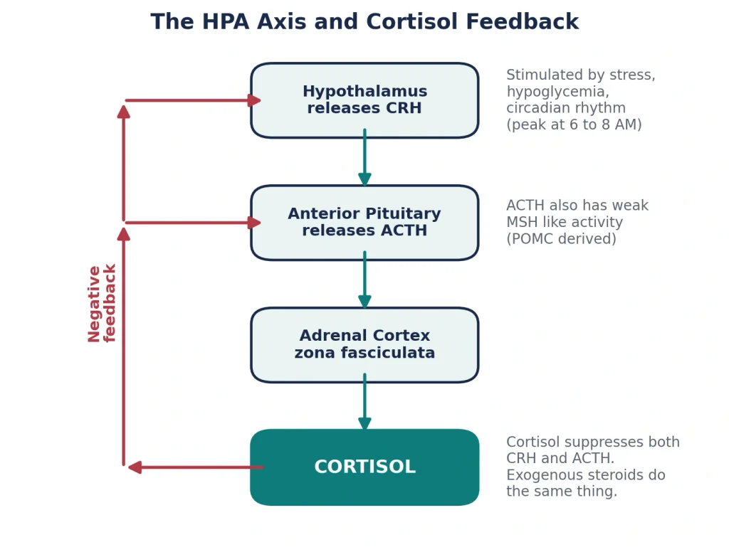
Figure 1. The HPA axis. Cortisol suppresses both CRH and ACTH. Exogenous steroids exploit the same feedback, which is why chronic steroid use shrinks the adrenal glands.
| ⚠️ Classic Trap Exogenous glucocorticoids suppress ACTH, so the adrenal glands atrophy bilaterally. A vignette of a patient on chronic prednisone will show LOW ACTH, LOW endogenous cortisol production, and small adrenals on imaging, even though the patient looks Cushingoid. Never pick adrenal hyperplasia for a patient on exogenous steroids. |
The adrenal gland is really two organs wrapped in one capsule. The cortex is mesoderm and makes steroids. The medulla is neural crest and makes catecholamines. The cortex itself has three zones, and the classic mnemonic works from outside in.
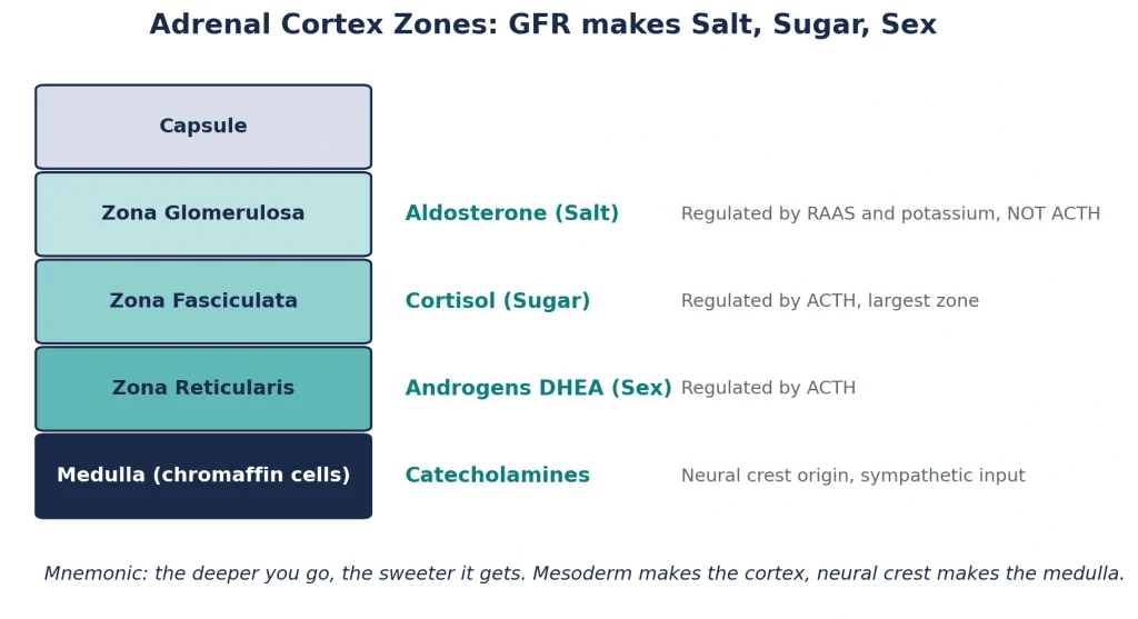
Figure 2. Adrenal cortex zones with their hormones and regulators. GFR from outside in, Salt Sugar Sex from superficial to deep, and the deeper you go the sweeter it gets.
Why does this zonation matter for our two diseases? Because it explains the single most tested difference between primary and secondary adrenal insufficiency. The glomerulosa listens to renin, not ACTH. So when the pituitary fails (secondary insufficiency), cortisol falls but aldosterone survives, and potassium stays normal. When the gland itself is destroyed (Addison disease), all three zones die together, aldosterone is lost, and the patient becomes hyperkalemic and salt wasting. This one anatomical fact resolves an entire family of exam questions.
You do not need every enzyme for this topic, but you need the skeleton because congenital adrenal hyperplasia questions share vignette space with Addison disease. Cholesterol enters the mitochondrion through the StAR protein, is converted to pregnenolone by desmolase, and then flows through 17 hydroxylase, 3 beta hydroxysteroid dehydrogenase, 21 hydroxylase, and 11 beta hydroxylase to become cortisol. ACTH upregulates the whole pathway, which is why chronic ACTH excess causes adrenal hyperplasia and chronic ACTH deficiency causes atrophy.
Aldosterone deserves its own paragraph because its independence from ACTH is the hinge on which many questions turn. Low renal perfusion triggers renin release from juxtaglomerular cells, renin converts angiotensinogen to angiotensin I, ACE converts that to angiotensin II, and angiotensin II plus hyperkalemia directly stimulate the zona glomerulosa. Aldosterone then acts on principal cells of the collecting duct to reabsorb sodium and secrete potassium, and on intercalated cells to secrete protons. Lose aldosterone and you lose sodium, retain potassium, and retain acid. That is the exact electrolyte triad of Addison disease: hyponatremia, hyperkalemia, and non anion gap metabolic acidosis.
ACTH is cut from a larger precursor called pro opiomelanocortin, and the same precursor yields melanocyte stimulating hormone. When ACTH rises to very high levels, its own weak MSH like activity plus co secreted MSH fragments stimulate melanocytes. This is the entire secret behind hyperpigmentation in Addison disease, and it is also why hyperpigmentation only appears when ACTH is high. Pituitary failure with low ACTH produces pale, not bronzed, patients. We will return to this repeatedly because the exam certainly will.
Image based adrenal questions are uncommon but predictable. The fasciculata appears as long cords of lipid rich, foamy clear cells. Autoimmune adrenalitis shows lymphocytic infiltration of the cortex with sparing of the medulla. Tuberculous adrenalitis shows caseating granulomas and often calcified, enlarged glands on CT. Metastases, classically from lung cancer, replace both glands and are one of the few causes of adrenal insufficiency with bilaterally enlarged adrenals. Embryologically, cortex from mesoderm and medulla from neural crest is a one line fact the exam recycles endlessly.
| 📌 Memorize This Adrenal insufficiency needs destruction of about 90 percent of both cortices before symptoms appear. This is why Addison disease presents insidiously with months of fatigue and weight loss, and why an acute stressor can suddenly unmask a gland that was quietly failing for a year. |
That’s the whole idea behind USMLE Impact Theory: learn one core mechanism, then recognize every disease that breaks it. Built by IMGs, for IMGs.
Cushing syndrome is the clinical state produced by chronic exposure to excess glucocorticoids from any source. The word syndrome is deliberately broad. It does not tell you where the cortisol came from. It only tells you the tissues have been soaked in cortisol long enough to change the body. The moment you see the word, your first job in any vignette is to ask one question: what is the ACTH doing
Students who memorize causes as a flat list get lost. Students who sort causes by ACTH answer in seconds. There are only four causes worth knowing, and the table below is the way we teach them: two with low ACTH, two with high ACTH.
| Cause | ACTH | Mechanism | Classic Vignette Clue |
|---|---|---|---|
| Exogenous glucocorticoids (most common overall) | Low | Oral, inhaled, injected, or topical steroids suppress the axis, bilateral adrenal atrophy | Patient with asthma, RA, IBD, or transplant on chronic prednisone |
| Adrenal adenoma or carcinoma | Low | Autonomous cortisol secretion suppresses ACTH, contralateral adrenal atrophies | Unilateral adrenal mass, carcinoma may also secrete androgens causing virilization |
| Cushing disease (pituitary ACTH adenoma) | High | Corticotroph adenoma drives bilateral adrenal hyperplasia, most common ENDOGENOUS cause | Young woman, gradual onset, suppresses with high dose dexamethasone |
| Ectopic ACTH secretion | High (very high) | Small cell lung cancer or bronchial carcinoid secretes ACTH with no feedback sensitivity | Smoker with weight LOSS, severe hypokalemia, hyperpigmentation, rapid onset |
| USMLE Pearl Three superlatives the exam loves: exogenous steroids are the most common cause of Cushing syndrome overall, Cushing disease is the most common endogenous cause, and small cell lung cancer is the most common source of ectopic ACTH. Read the question stem carefully to see which superlative is being asked. |
Every clinical feature of Cushing syndrome is a direct extension of normal cortisol physiology turned up too far. Cortisol is catabolic in the periphery and anabolic for glucose. Walk through the tissues one by one and the entire syndrome writes itself.
| ⚠️ Classic Trap Severe hypokalemia with alkalosis points to ectopic ACTH, not Cushing disease. Ectopic tumors produce cortisol levels so extreme that renal 11 beta HSD2 is saturated and cortisol floods the mineralocorticoid receptor. A smoker with hypokalemia of 2.8 and hyperglycemia is small cell lung cancer until proven otherwise. |
The exam presents Cushing syndrome either as a classic picture or as a deliberately incomplete one. This table separates the findings that are most specific from the ones that are common but nonspecific, which is exactly how you should weigh them in a vignette.
| Most Specific Findings | Common but Less Specific Findings |
|---|---|
| Wide purple striae greater than 1 cm | Central obesity and weight gain |
| Proximal muscle weakness (difficulty rising from a chair) | Hypertension |
| Easy bruising and thin skin | Glucose intolerance |
| Facial plethora | Depression, irritability, insomnia |
| Osteoporosis in a young patient | Menstrual irregularity, hirsutism, acne |
A useful clinical rule that also works on the exam: obesity and hypertension are everywhere, but thin skin, proximal weakness, and unexplained fractures in a young person are the findings that should force Cushing syndrome to the top of your differential.
Diagnosis always proceeds in the same two steps, and the exam punishes anyone who does them out of order. Step one, prove hypercortisolism exists. Step two, localize its source with ACTH. Never order a pituitary MRI before you have confirmed cortisol excess biochemically, because incidental pituitary lesions are common enough to mislead you.
Three screening tests establish hypercortisolism, and any of them can appear in a stem.
Once hypercortisolism is confirmed, a single ACTH measurement splits the world in two. Low ACTH means the problem is adrenal or exogenous, so image the adrenals with CT. High ACTH means the problem is pituitary or ectopic, so proceed to the high dose dexamethasone test or inferior petrosal sinus sampling, then image accordingly.
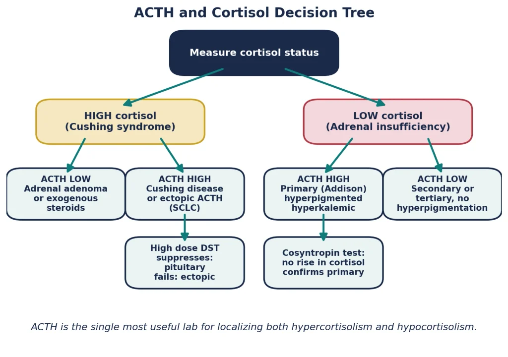
Figure 3. The ACTH and cortisol decision tree. One hormone pair localizes essentially every adrenal axis disorder on the exam.
Dexamethasone is a synthetic glucocorticoid that suppresses ACTH in any tissue that still respects feedback. The test simply asks each possible source of cortisol: do you still listen to feedback at all
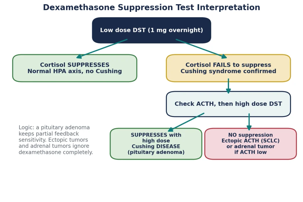
Figure 4. Interpreting low dose and high dose dexamethasone suppression, the single most tested algorithm in adrenal endocrinology.
Here is the same information as a rapid interpretation table you can photograph with your mind the night before the exam.
| Condition | ACTH | Low Dose DST | High Dose DST |
|---|---|---|---|
| Normal person | Normal | Suppresses | Suppresses |
| Cushing disease (pituitary) | High | No suppression | Suppresses |
| Ectopic ACTH (SCLC, carcinoid) | Very high | No suppression | No suppression |
| Adrenal adenoma or carcinoma | Low | No suppression | No suppression |
| Exogenous steroids | Low | Cortisol already low | Cortisol already low |
| Examiner’s Favorite When high dose dexamethasone fails to suppress and ACTH is high, the next best test is inferior petrosal sinus sampling, which directly compares central versus peripheral ACTH. A central to peripheral gradient confirms the pituitary. This is the classic next step question for ambiguous cases. |
Book a free call with mentors who matched as IMGs. We’ll build a plan that untangles every high-yield trap the same way, one clear mechanism at a time.
Imaging comes last and follows the biochemistry. Low ACTH sends you to a CT of the adrenal glands looking for a unilateral mass with contralateral atrophy. High ACTH that is suppressed with high dose dexamethasone sends you to a pituitary MRI looking for a microadenoma, remembering that many corticotroph adenomas are small enough to hide. High ACTH that does not suppress sends you to a CT of the chest hunting for small cell lung cancer or a bronchial carcinoid. Ordering imaging in this sequence is itself a tested skill.
Treatment is satisfying because it maps one to one onto the cause. The table below covers every therapeutic fact Step 1 and Step 2 CK realistically test, including the perioperative trap that follows successful surgery.
| Cause | First Line Treatment | Key Additional Points |
|---|---|---|
| Exogenous steroids | Gradual taper of the steroid | Never stop abruptly, the atrophied adrenals cannot respond and adrenal crisis follows |
| Cushing disease | Transsphenoidal resection of the pituitary adenoma | Pasireotide, cabergoline, or bilateral adrenalectomy for refractory cases, watch for Nelson syndrome after adrenalectomy |
| Adrenal adenoma or carcinoma | Adrenalectomy | Mitotane is adrenolytic for carcinoma, contralateral gland is suppressed so cover with steroids postoperatively |
| Ectopic ACTH | Resect or treat the underlying tumor | Ketoconazole, metyrapone, or etomidate to block cortisol synthesis when the tumor cannot be controlled |
| ⚠️ Never Confuse After curative surgery for any endogenous Cushing syndrome, the remaining normal axis has been suppressed for months and is asleep. Patients need stress dose steroids and a slow taper postoperatively or they will present with adrenal insufficiency days after their cure. The exam adores this irony. |
The terminology trips up more students than the physiology, so let us be exact. Cushing syndrome is the state of cortisol excess from any cause. Cushing disease is one specific cause: an ACTH secreting adenoma of the anterior pituitary. Every patient with Cushing disease has Cushing syndrome, but only a minority of Cushing syndrome is Cushing disease. On imaging, Cushing disease shows a pituitary microadenoma with bilateral adrenal hyperplasia, in contrast to the unilateral mass of an adrenal adenoma. Demographically it favors women between 20 and 40. Biochemically it is the only high ACTH cause that suppresses high dose dexamethasone. Treatment is transsphenoidal surgery, and if both adrenals are ever removed instead, the unopposed adenoma can enlarge aggressively and pigment the skin, a complication called Nelson syndrome.
| Step 1 High Yield Vignette translation drill: Cushingoid appearance plus bilateral adrenal enlargement means the ACTH is high and the driver is above the adrenals. Cushingoid appearance plus a unilateral adrenal mass with an atrophic partner means the adrenal itself is the driver and ACTH is low. Let the adrenal anatomy tell you the ACTH before the labs do. |
Chronic alcohol use, severe obesity, poorly controlled diabetes, and major depression can all mildly elevate cortisol and blur screening tests. The exam occasionally gestures at this with an alcohol vignette. The practical points are that pseudo Cushing states usually retain diurnal rhythm, improve when the underlying condition is treated, and lack the specific catabolic signs such as wide purple striae and proximal myopathy. When screening results conflict, repeat testing and the dexamethasone CRH test help separate the two.
Addison disease is primary adrenal insufficiency, meaning the adrenal cortex itself is destroyed and can no longer make adequate cortisol, aldosterone, or adrenal androgens. Because the failure is in the gland, the pituitary responds the only way it can, by shouting louder. ACTH climbs relentlessly, and that single hormonal fact generates the most photogenic finding in endocrinology, the bronzed skin of Addison disease. Contrast this from the start with secondary insufficiency, where the pituitary itself has failed, ACTH is low, aldosterone is preserved, and the skin is pale.
The causes divide neatly by geography and by tempo, and both dimensions appear in vignettes. The table below organizes them the way examiners think.
| Cause | Key Features and Vignette Clues |
|---|---|
| Autoimmune adrenalitis | Most common cause in the developed world, antibodies against 21 hydroxylase, associated with other autoimmune disease such as Hashimoto thyroiditis, type 1 diabetes, vitiligo, and the autoimmune polyendocrine syndromes |
| Tuberculosis | Most common cause worldwide in endemic regions, enlarged then calcified adrenals on CT, immigrant or endemic exposure history |
| Bilateral adrenal metastases | Lung cancer classically, one of the few insufficiency causes with big adrenals |
| Waterhouse Friderichsen syndrome | Bilateral hemorrhagic adrenal infarction in overwhelming sepsis, classically Neisseria meningitidis |
| Adrenal hemorrhage or infarction | Anticoagulation, trauma, antiphospholipid syndrome, DIC |
| Infections in HIV | CMV, disseminated fungal infection such as histoplasmosis |
| Drugs | Ketoconazole and etomidate block synthesis, rifampin accelerates cortisol metabolism, abrupt steroid withdrawal unmasks a suppressed axis |
| Congenital adrenal hyperplasia | 21 hydroxylase deficiency in a neonate with salt wasting and virilization |
Addison disease is famously insidious. The symptoms are vague for months, which is exactly why the exam trains you to catch the specific combination rather than any single complaint.
Notice how the picture is a photographic negative of Cushing syndrome. Weight loss instead of central obesity, hypotension instead of hypertension, hypoglycemia instead of hyperglycemia, pigmented thin skin instead of pale thin skin with striae. Learning the two diseases as mirror images halves the memory load.
This mechanism is asked so often that it deserves its own section. ACTH is cleaved from pro opiomelanocortin, and POMC processing also yields melanocyte stimulating hormone. ACTH itself shares sequence homology with MSH and weakly stimulates the melanocortin 1 receptor on melanocytes. In Addison disease, cortisol feedback is gone, POMC transcription runs uncontrolled, and ACTH with its MSH like fragments rises high enough to activate melanocytes throughout the skin and mucosa. The result is diffuse bronzing concentrated where melanocytes are most active, in creases, friction areas, scars, and gums.
| 📌 Memorize This Hyperpigmentation is an ACTH story, not a cortisol story. It appears wherever ACTH is chronically high: Addison disease, ectopic ACTH from small cell lung cancer, and Nelson syndrome. It never appears in secondary adrenal insufficiency or in adrenal adenoma, because in both of those ACTH is low. |
Every abnormal value in the Addison lab panel is traceable to one of the two missing hormones. Aldosterone loss means the collecting duct stops reabsorbing sodium and stops secreting potassium and protons, giving hyponatremia, hyperkalemia, and a non anion gap metabolic acidosis with volume depletion. Cortisol loss contributes its own hyponatremia because low cortisol disinhibits ADH release and impairs free water excretion. Cortisol loss also produces hypoglycemia from lost gluconeogenesis, and the chronically high ACTH does the pigmenting. A modest eosinophilia rounds out the classic panel, since cortisol normally suppresses eosinophils.
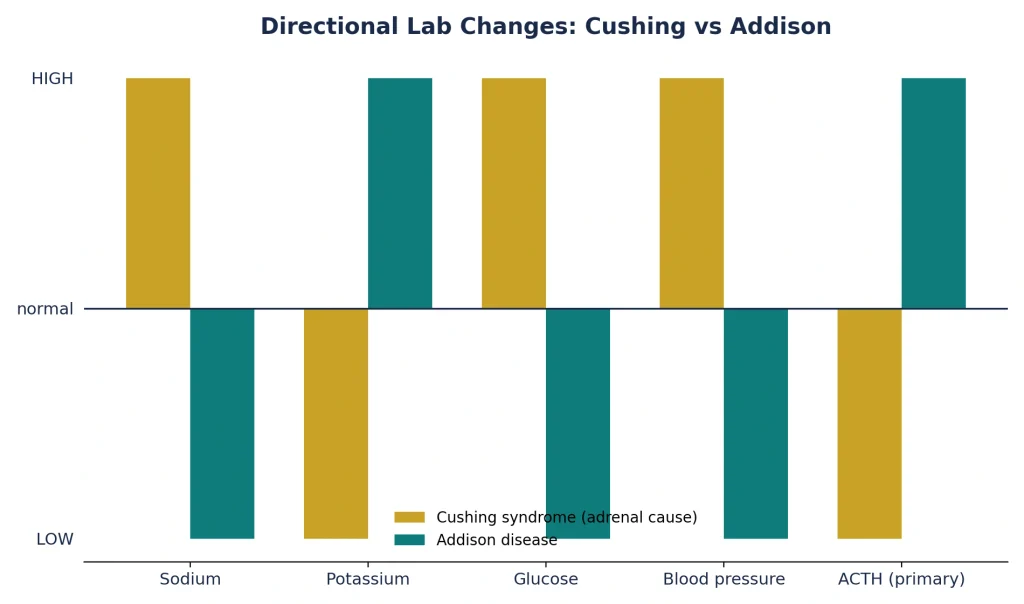
Figure 5. Directional lab changes in Cushing syndrome versus Addison disease. The two diseases are mirror images across almost every axis.
The workup answers two questions in order, exactly parallel to Cushing syndrome. First, is cortisol truly deficient. Second, is the lesion in the gland or above it. A morning cortisol that is very low with a simultaneously high ACTH essentially makes the diagnosis of primary insufficiency. When results are equivocal, the cosyntropin stimulation test is the confirmatory step: give synthetic ACTH and measure cortisol response. A destroyed gland cannot respond no matter how loudly it is stimulated, so cortisol fails to rise in Addison disease. In chronic secondary insufficiency the atrophic but intact gland also responds poorly, so the ACTH level, not the stimulation test alone, is what localizes the lesion. Checking renin and aldosterone, 21 hydroxylase antibodies, and a chest evaluation for TB completes the picture.
| Test | Primary (Addison) | Secondary (Pituitary) |
|---|---|---|
| Morning cortisol | Low | Low |
| ACTH | High | Low or inappropriately normal |
| Cosyntropin stimulation | No adequate rise | Blunted rise in chronic disease |
| Aldosterone and renin | Aldosterone low, renin high | Aldosterone preserved |
| Potassium | High | Normal |
| Hyperpigmentation | Present | Absent |
The exam frames this comparison in a dozen costumes, but it is always the same three door puzzle. Primary means the gland failed. Secondary means the pituitary failed, from a mass, surgery, radiation, Sheehan syndrome, or apoplexy. Tertiary means the hypothalamus is suppressed, and its overwhelmingly common cause is chronic exogenous steroid use that is stopped abruptly. Secondary and tertiary behave alike: low ACTH, no hyperpigmentation, normal potassium because aldosterone still answers to renin, though hyponatremia can still occur through cortisol dependent water handling. If a vignette gives you a hypotensive patient whose potassium is normal and whose skin is pale, look upward, not at the gland.
| USMLE Pearl The fastest discriminator in any adrenal insufficiency vignette is the pairing of potassium and skin. High potassium plus dark skin equals primary. Normal potassium plus pale skin equals secondary or tertiary. Two data points, one diagnosis. |
Treatment replaces what was lost, for life. Hydrocortisone in divided doses mimics the diurnal rhythm and replaces glucocorticoid. Fludrocortisone replaces mineralocorticoid and corrects the sodium, potassium, and volume problems. Some women benefit from DHEA replacement. The teaching that saves lives, and that the exam tests as sick day rules, is that patients must increase their glucocorticoid dose during febrile illness, vomiting, trauma, or surgery, and must carry injectable hydrocortisone and medical identification, because the one thing an Addisonian patient cannot do is mount a stress response.
Adrenal crisis is acute, life threatening adrenal insufficiency. It appears in a patient with known Addison disease hit by infection or surgery, in an undiagnosed patient unmasked by stress, in abrupt withdrawal of chronic steroids, or in acute hemorrhagic destruction of the glands. The presentation is shock that behaves wrongly: hypotension refractory to fluids and vasopressors, with vomiting, abdominal pain sometimes rigid enough to mimic a surgical abdomen, fever, hypoglycemia, hyponatremia, hyperkalemia, and altered mental status.
Management is a reflex, not a deliberation, and the order of operations is itself the exam answer. Draw cortisol and ACTH, then treat immediately without waiting: IV hydrocortisone 100 mg plus aggressive saline and dextrose, then hunt for and treat the precipitant. High dose hydrocortisone has enough intrinsic mineralocorticoid activity that fludrocortisone can wait until maintenance dosing.
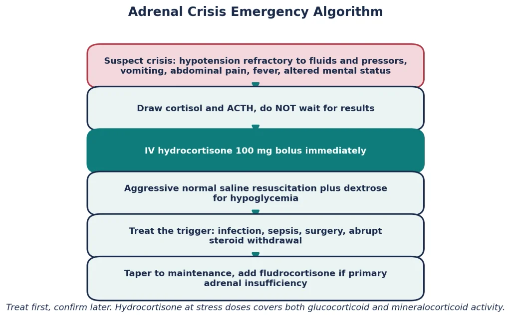
Figure 6. Adrenal crisis management. Treat first and confirm later, because the confirmatory labs survive treatment but the patient may not survive the delay.
| ⚠️ Never Confuse In a vignette of refractory shock, checking cortisol is wise but waiting for the result before giving hydrocortisone is the wrong answer. If dexamethasone is used instead of hydrocortisone, it has the advantage of not interfering with the cosyntropin test, a nuance Step 2 CK enjoys. |
Waterhouse Friderichsen syndrome is fulminant adrenal failure from bilateral hemorrhagic infarction of the adrenal glands during overwhelming septicemia. The classic organism is Neisseria meningitidis, though pneumococcus and other severe infections can do the same. The vignette is unforgettable once seen: a child or young adult with an abrupt fever, a spreading petechial or purpuric rash, DIC, and catastrophic hypotension collapsing over hours. The endotoxin driven coagulopathy bleeds into the highly vascular adrenals and destroys them acutely, so there has been no time for hyperpigmentation. Treatment is immediate antibiotics, stress dose steroids, and intensive care, and mortality is high even when everything is done right.
Conn syndrome is primary hyperaldosteronism, usually from an aldosterone producing adenoma of the zona glomerulosa or from bilateral idiopathic hyperplasia. It matters here because examiners love to hide it among Cushing and Addison options. The picture is hypertension that is resistant to therapy, hypokalemia with muscle weakness or cramps, metabolic alkalosis, and a suppressed renin. Sodium sits high normal without dramatic edema because of aldosterone escape. Screening is an aldosterone to renin ratio, confirmation is failure to suppress aldosterone with salt loading, and treatment is adrenalectomy for adenoma or a mineralocorticoid receptor antagonist such as spironolactone or eplerenone for hyperplasia. The three way comparison below is one of the highest value tables in this entire guide.
| Feature | Cushing Syndrome | Conn Syndrome | Addison Disease |
|---|---|---|---|
| Hormone problem | High cortisol | High aldosterone | Low cortisol and aldosterone |
| Blood pressure | High | High | Low |
| Potassium | Normal or low | Low | High |
| Acid base | Alkalosis when severe | Metabolic alkalosis | Metabolic acidosis |
| Glucose | High | Normal | Low |
| Renin | Variable | Suppressed | High |
| Distinct clues | Striae, moon facies, proximal weakness | Refractory hypertension, no Cushingoid features | Hyperpigmentation, weight loss, salt craving |
Both diseases have impostors, and question writers use them as distractors. This table lists the differentials worth knowing and the single feature that separates each from the real thing.
| Presentation | Mimic | How to Tell Them Apart |
|---|---|---|
| Cushingoid appearance | Metabolic syndrome and simple obesity | No proximal weakness, no wide purple striae, normal dexamethasone suppression |
| Cushingoid appearance | Pseudo Cushing of alcohol use or depression | Diurnal rhythm preserved, resolves with abstinence or treatment |
| Cushingoid appearance | Polycystic ovary syndrome | Hirsutism and irregular menses without catabolic skin and muscle findings |
| Fatigue with weight loss | Hyperthyroidism, malignancy, chronic infection | No hyperkalemia, no hyperpigmentation, cortisol axis normal |
| Hyperpigmentation | Hemochromatosis | Diabetes and liver disease with high ferritin, ACTH normal |
| Hyperkalemia with acidosis | Type 4 renal tubular acidosis | Hyporeninemic hypoaldosteronism of diabetic kidney disease, cortisol normal |
| Shock with abdominal pain | Surgical abdomen or sepsis alone | Refractory hypotension responding dramatically to hydrocortisone suggests adrenal crisis |
Work through these actively. Read the stem, commit to a diagnosis and a next step, then read the answer. Each case is built from a pattern that has appeared in NBME material in some form.
A 34 year old woman has 8 kilograms of weight gain, facial rounding, and new hypertension over one year. Wide violaceous striae cross her abdomen and she cannot rise from a squat. Late night salivary cortisol is elevated twice, and ACTH is high. High dose dexamethasone suppresses her cortisol. Answer: Cushing disease from a pituitary microadenoma. Next step is pituitary MRI, then transsphenoidal resection.
A 61 year old man with a 45 pack year history has lost 10 kilograms, and has potassium of 2.7, glucose of 240, darkening knuckles, and muscle weakness that developed over six weeks. ACTH is markedly elevated and high dose dexamethasone changes nothing. Answer: ectopic ACTH from small cell lung cancer. Weight loss instead of gain, speed, and severe hypokalemia are the giveaways. Next step is chest CT.
A 28 year old woman with vitiligo reports a year of fatigue, salt craving, nausea, and a 7 kilogram weight loss. She is hypotensive with darkened palmar creases and gingival pigmentation. Sodium is 128, potassium 5.9. Answer: autoimmune Addison disease. Confirm with morning cortisol plus ACTH and a cosyntropin test, then start hydrocortisone and fludrocortisone.
A 45 year old man on 20 mg of prednisone daily for three years for rheumatoid arthritis stops it abruptly before a dental procedure. Two days later he is hypotensive, vomiting, and hypoglycemic. Potassium is normal and his skin is pale. Answer: tertiary adrenal insufficiency from abrupt steroid withdrawal precipitating adrenal crisis. Aldosterone is intact, hence the normal potassium. Give IV hydrocortisone immediately.
A 19 year old college student develops fever, headache, and a rapidly spreading purpuric rash, then collapses with a blood pressure of 60 over 30 that does not respond to three liters of saline and norepinephrine. Answer: Waterhouse Friderichsen syndrome complicating meningococcemia. Immediate ceftriaxone plus IV hydrocortisone, expect DIC on labs.
A 52 year old woman has an incidental 2.5 centimeter left adrenal mass. She has hypertension, easy bruising, and thin skin. ACTH is undetectable and low dose dexamethasone fails to suppress cortisol. The right adrenal appears atrophic. Answer: cortisol secreting adrenal adenoma. Adrenalectomy with perioperative steroid coverage, because the suppressed right gland cannot yet function.
A 39 year old woman treated with transsphenoidal surgery is cured of Cushing disease. Five days after discharge she presents with fatigue, nausea, and orthostatic hypotension. Answer: postoperative secondary adrenal insufficiency, the suppressed normal corticotrophs have not recovered. She needed a maintenance steroid taper. Treat with hydrocortisone.
A 47 year old man has hypertension on four drugs, potassium of 3.0, and metabolic alkalosis. He has no striae, no weakness, and normal glucose. Renin is suppressed and the aldosterone to renin ratio is high. Answer: Conn syndrome. Confirm with salt suppression testing, then adrenal CT and adrenal vein sampling to distinguish adenoma from bilateral hyperplasia.
A 33 year old woman with postpartum hemorrhage one year ago has failed to lactate, has amenorrhea, cold intolerance, and now fatigue with hypotension. Potassium is normal, skin is pale, ACTH and cortisol are both low. Answer: Sheehan syndrome causing secondary adrenal insufficiency within panhypopituitarism. Replace cortisol before thyroid hormone to avoid precipitating crisis.
A 6 day old newborn presents with vomiting, dehydration, sodium of 126, potassium of 6.8, and ambiguous genitalia was noted in the female infant at birth. Answer: salt wasting congenital adrenal hyperplasia from 21 hydroxylase deficiency, biochemically an adrenal crisis. Elevated 17 hydroxyprogesterone confirms it. Treat with fluids, hydrocortisone, and fludrocortisone.
USMLE Impact Theory covers every high-yield topic the same way, NBME-style cases, wrong-answer analysis, and the exact traps written into the stems.
Mnemonics work best when they encode logic, not just letters. Each of the following was written for this guide, and each line is worth expanding back into the physiology it compresses.
After mentoring thousands of IMGs through Step 1, we can tell you that adrenal questions cluster into a small number of repeating formats. Recognize the format and you have half the answer.
Adrenal disorders show up on almost every Step 1 form, usually as an ACTH-and-cortisol reasoning question rather than simple recall. That is why they sit on nearly every list of high-yield Step 1 topics, one vignette can test physiology, pathology, and pharmacology at once. Learn to read the hormone arrows and the answer usually falls out.
| Question Format | What It Really Tests | Your Anchor |
|---|---|---|
| Vignette then asks for ACTH and cortisol arrows | Localization along the axis | Draw the three story axis, break the level described, read off the arrows |
| Dexamethasone results given, asks diagnosis | Feedback sensitivity logic | Only pituitary adenomas suppress with high dose |
| Electrolyte panel, asks the hormone problem | Aldosterone physiology | High potassium means the gland is dead, normal potassium means the pituitary is |
| Skin pigmentation, asks the mechanism | POMC biochemistry | ACTH and MSH share a precursor |
| Refractory shock, asks next step | Adrenal crisis recognition | Hydrocortisone now, confirmation later |
| Smoker with hypokalemia, asks the source | Ectopic ACTH pattern | Small cell lung cancer, weight loss not gain |
| Post surgical steroid question | Axis suppression and recovery | Taper always, stress dosing for illness and surgery |
| Buzzword image of striae or bronzing | Pattern recognition | Wide purple striae for Cushing, creases and gums for Addison |
| USMLE Pearl Frequently confused pairs to rehearse the night before: Cushing disease vs Cushing syndrome, primary vs secondary insufficiency, ectopic ACTH vs Cushing disease, Conn vs Cushing hypokalemia, hemochromatosis bronzing vs Addison bronzing, and adrenal crisis vs septic shock. If you can articulate the one line difference for each pair out loud, you are ready. |
A guide this size is a reference, not a one-sitting read. Come back to this revision sheet the week before your exam, and fit focused reps like it into your daily plan, our breakdown of how many hours a day to study for Step 1 helps you budget the endocrine block realistically.
Cushing syndrome is the collection of body changes caused by too much cortisol from any source, whether steroid medication, an adrenal tumor, a pituitary tumor, or an ACTH producing cancer. The hallmark features are central weight gain, moon facies, purple striae, hypertension, high glucose, and muscle weakness.
Addison disease is failure of the adrenal cortex itself, so the body lacks both cortisol and aldosterone. Patients develop fatigue, weight loss, low blood pressure, salt craving, low sodium, high potassium, and darkening of the skin driven by high ACTH.
Cushing syndrome is the general state of cortisol excess. Cushing disease is one specific cause of that state, an ACTH secreting pituitary adenoma. All Cushing disease is Cushing syndrome, but most Cushing syndrome, especially steroid induced, is not Cushing disease.
Because ACTH and melanocyte stimulating hormone come from the same precursor molecule, POMC. When cortisol feedback disappears, ACTH production rises massively, and its MSH like activity stimulates melanocytes, bronzing the skin, creases, scars, and gums.
In secondary insufficiency the pituitary itself has failed, so ACTH is low. Without high ACTH there is no melanocyte stimulation, and the patient is pale rather than bronzed. Pigmentation is a marker of high ACTH, not of low cortisol.
Potassium is controlled by aldosterone, and aldosterone is controlled by renin rather than ACTH. Addison disease destroys the whole cortex including the aldosterone producing zona glomerulosa, so potassium rises. In secondary insufficiency the gland is intact and renin keeps aldosterone running, so potassium stays normal.
Dexamethasone is a synthetic steroid that suppresses ACTH in a normal, feedback sensitive axis. In a healthy person 1 mg at night suppresses the next morning cortisol. In any true Cushing syndrome the cortisol fails to suppress, which makes the low dose test a screening tool for the presence of hypercortisolism.
A pituitary adenoma retains partial feedback sensitivity, so a large enough dexamethasone dose finally suppresses its ACTH and cortisol. Ectopic tumors such as small cell lung cancer carry no feedback machinery, so even high doses change nothing. Suppression points to the sella, no suppression points to the chest.
High ACTH occurs in Cushing disease, ectopic ACTH secretion, and Addison disease. Low ACTH occurs in adrenal adenoma, exogenous steroid use, and secondary adrenal insufficiency. Whether cortisol is high or low then tells you which of the two matching conditions you are in.
Exogenous glucocorticoid therapy is the most common cause overall. Among endogenous causes, Cushing disease from a pituitary adenoma is the most common, and among ectopic sources, small cell lung cancer leads.
Simulate exam conditions: cover the explanation, commit to an answer, and only then read on. Each question includes the reasoning for the right answer and against every wrong one, because reviewing distractors is where the real learning happens.
A 36 year old woman presents with 10 kilograms of weight gain, facial rounding, and secondary amenorrhea over 14 months. Examination shows abdominal striae 1.5 centimeters wide, proximal muscle weakness, and a blood pressure of 158 over 98. Late night salivary cortisol is elevated on two occasions. Serum ACTH is elevated. Cortisol suppresses after high dose dexamethasone. Which of the following is the most likely diagnosis
Correct answer: C. Confirmed hypercortisolism with a high ACTH narrows the field to pituitary versus ectopic, and suppression with high dose dexamethasone identifies the pituitary, which retains partial feedback sensitivity. A is wrong because an adrenal adenoma suppresses ACTH. B is wrong because ectopic tumors do not suppress with any dexamethasone dose. D is wrong because exogenous steroids produce low ACTH. E is wrong because cortisol is high, not low. Learning objective: localize hypercortisolism using ACTH and dexamethasone responses. Difficulty: moderate. High yield takeaway: suppression says sella.
A 63 year old man with a 50 pack year smoking history presents with 8 kilograms of weight loss over two months, new hyperglycemia, and profound weakness. Blood pressure is 165 over 100, potassium is 2.6, and bicarbonate is 34. Skin creases appear darkened. Which of the following is the most likely source of his condition
Correct answer: B. Rapid onset, weight loss rather than gain, severe hypokalemic alkalosis, and hyperpigmentation mark extreme ACTH from an ectopic source, and small cell lung cancer in a heavy smoker is the classic culprit. A produces a slower, milder syndrome with weight gain. C would suppress ACTH and cause no pigmentation. D causes cortisol deficiency, the opposite picture. E lowers ACTH and cannot pigment the skin. Learning objective: recognize the ectopic ACTH phenotype. Difficulty: moderate. High yield takeaway: Cushing with weight loss and potassium in the twos is ectopic until proven otherwise.
A 29 year old woman with Hashimoto thyroiditis reports a year of fatigue, nausea, salt craving, and a 6 kilogram weight loss. Blood pressure is 88 over 56 with orthostasis. Pigmentation is noted along palmar creases and the buccal mucosa. Laboratory studies show sodium 127, potassium 6.0, and glucose 62. Which set of findings is most likely
Correct answer: B. Autoimmune destruction of the whole cortex lowers cortisol and aldosterone together, and lost feedback drives ACTH high, which also explains the pigmentation. A and D describe secondary insufficiency, which spares aldosterone and cannot pigment. C describes no coherent disease. E is physiologically inconsistent because low cortisol should raise ACTH. Learning objective: predict the full hormone panel of primary adrenal insufficiency. Difficulty: easy. High yield takeaway: pigment plus hyperkalemia equals primary disease with high ACTH.
A 41 year old man with severe asthma has taken high dose inhaled and frequent oral corticosteroids for four years. He develops moon facies and easy bruising. Which additional finding is most likely on evaluation
Correct answer: C. Exogenous steroids suppress CRH and ACTH, and without trophic stimulation both adrenal glands atrophy. A requires high ACTH. B is the opposite of what feedback produces. D requires high ACTH. E is a subtle trap: urinary free cortisol measures endogenous cortisol, which is suppressed, while many synthetic steroids are not detected as cortisol. Learning objective: understand the axis consequences of exogenous glucocorticoids. Difficulty: moderate. High yield takeaway: iatrogenic Cushing runs on a suppressed, atrophied axis.
A 19 year old man is brought to the emergency department with fever, headache, and a petechial rash that has spread over four hours. Blood pressure is 62 over 34 despite three liters of crystalloid and escalating norepinephrine. Laboratory studies show thrombocytopenia, prolonged PT and PTT, and elevated D dimer. Which intervention addresses the most likely endocrine complication
Correct answer: B. Meningococcemia with DIC and refractory shock suggests Waterhouse Friderichsen syndrome, hemorrhagic destruction of both adrenals, and the lifesaving move is empiric stress dose hydrocortisone alongside antibiotics. A is a diagnostic test for cortisol excess, irrelevant here. C lacks the needed glucocorticoid activity and is not the acute drug. D removes what is already destroyed. E is the classic fatal delay. Learning objective: manage fulminant adrenal failure in sepsis. Difficulty: easy. High yield takeaway: refractory shock plus purpura means steroids now.
A 55 year old woman with an incidentally discovered 3 centimeter right adrenal mass has hypertension, thin skin, and new diabetes. ACTH is undetectable and cortisol fails to suppress with 1 mg of dexamethasone. She undergoes right adrenalectomy. Which perioperative complication is she at greatest risk for
Correct answer: C. Her autonomous adenoma suppressed ACTH for years, so the left adrenal is atrophic and cannot secrete cortisol the moment the tumor is removed. Without perioperative steroid coverage she becomes acutely insufficient. A follows bilateral adrenalectomy in Cushing disease, a different setting. B and D involve hormones her lesion did not secrete. E is unrelated to the operation. Learning objective: anticipate axis suppression after curing cortisol excess. Difficulty: hard. High yield takeaway: curing Cushing creates temporary Addison, cover with steroids and taper.
A 44 year old woman treated with chronic high dose prednisone for lupus nephritis stops all medications on her own. Three days later she presents with hypotension, vomiting, and a glucose of 48. Serum sodium is 129 and potassium is 4.1. Which feature best explains her normal potassium
Correct answer: A. Steroid withdrawal causes central, tertiary insufficiency. The gland is structurally intact, and since the glomerulosa answers to renin and potassium rather than ACTH, aldosterone continues and potassium stays normal. B is imprecise, cortisol has minor effects but the real answer is preserved aldosterone. C inverts physiology, low ACTH does not stimulate anything. D is false, any primary destruction causes hyperkalemia. E misunderstands prednisone, which has little mineralocorticoid effect and is now stopped. Learning objective: separate central from primary insufficiency using potassium. Difficulty: moderate. High yield takeaway: normal potassium in adrenal insufficiency points above the gland.
A 50 year old man has hypertension requiring four medications. Potassium is 2.9 and bicarbonate is 32. He has no striae, normal glucose, normal body habitus, and no proximal weakness. Plasma renin activity is suppressed and the aldosterone to renin ratio is markedly elevated. Which of the following is the most likely diagnosis
Correct answer: C. Resistant hypertension with hypokalemic metabolic alkalosis and a suppressed renin defines Conn syndrome. A and B cause cortisol stigmata and, in ectopic disease, hyperglycemia with weight change, which are absent. D produces the opposite pattern of hypotension and hyperkalemia. E causes secondary hyperaldosteronism with HIGH renin, the single lab that separates it. Learning objective: use renin to classify hyperaldosteronism. Difficulty: moderate. High yield takeaway: suppressed renin turns hypertension with hypokalemia into Conn syndrome.
A 31 year old woman is evaluated for fatigue. Morning cortisol is low. Cosyntropin administration produces a robust rise in cortisol. Serum ACTH is low. Brain MRI shows a pituitary macroadenoma. Which additional finding is most consistent with her adrenal axis lesion
Correct answer: C. Low cortisol with low ACTH and a responsive gland defines secondary adrenal insufficiency from the pituitary mass. Aldosterone remains renin driven, so potassium is normal, and without high ACTH there is no pigmentation, so the patient is pale. A and B belong to primary disease. D describes primary aldosterone failure, not present here. E is the primary pattern of mineralocorticoid loss. Learning objective: build the complete secondary insufficiency phenotype. Difficulty: moderate. High yield takeaway: secondary insufficiency is pale, normokalemic, and low in ACTH.
A 27 year old woman with known Addison disease on hydrocortisone and fludrocortisone develops influenza with vomiting and cannot keep her medications down. She arrives confused with a blood pressure of 74 over 40, glucose 44, sodium 126, and potassium 6.1. After drawing blood for cortisol and ACTH, which is the best immediate management
Correct answer: C. This is adrenal crisis precipitated by illness and missed doses. Immediate parenteral hydrocortisone with volume and glucose replacement is the standard, and at stress doses hydrocortisone covers mineralocorticoid needs too. A fails because she cannot absorb oral drugs while vomiting. B repeats the classic fatal delay. D addresses electrolytes without fixing the cortisol emergency. E will underperform because shock in cortisol deficiency resists catecholamines until steroids are replaced. Learning objective: execute adrenal crisis management in the correct order. Difficulty: easy. High yield takeaway: salt, sugar, steroids, search.
If this guide did its job, Cushing syndrome and Addison disease no longer feel like two lists of findings but like one axis you can bend in either direction and read like an instrument panel. That shift, from memorizing diseases to modeling systems, is the entire philosophy behind how we teach at IMG Helping Hands, and it is the difference between students who survive the endocrine section and students who look forward to it. Keep the rapid revision sheet for exam week, redo the ten questions until every distractor feels transparent, and teach the hyperpigmentation mechanism to a friend, because the concept you can explain is the concept you will never lose. We will see you in the next guide.
This is exactly how USMLE Impact Theory works, built by IMGs, for IMGs, on the same physiology-first engine you just used to separate two diseases everyone confuses. Here’s what that looks like across the exam:
Articles published by IMG Helping Hands are prepared by our team using information from direct experience, publicly available resources, and educational references. AI tools may be used to assist with drafting, proofreading, and formatting; however, all content undergoes review and approval before publication.
The information provided is intended for educational purposes only. Requirements, policies, and processes may change over time. Readers should consult official sources for the most current information.