

Every IMG who has sat down with First Aid open to the renal chapter knows this feeling. Two syndromes. Almost identical names. Overlapping diseases. Buzzwords that look interchangeable. A new vocabulary of casts, immunofluorescence patterns, and electron microscopy findings that all start to blur by the second hour of study.
Here is the truth nobody tells you. You are not confused because nephrology is hard. You are confused because most resources teach you to memorize before you understand the pattern. At IMG Helping Hands, our UIT (Ultimate Integrated Teaching) approach flips this. We teach the mechanism first, the pattern second, and the buzzwords last. When you finish this guide, nephrotic and nephritic will feel as different as pneumonia and asthma. Step 1 vignettes will solve themselves.
This is the guide we wish we had when we were studying. Save it. Revisit it before your exam. Share it with the IMG next to you who is drowning in renal pathology.
| Who This Guide is For Every IMG preparing for USMLE Step 1. Every Step 2 CK candidate who needs to refresh renal pathology. Every clinical student who wants to walk into rounds knowing what an RBC cast actually means. Every educator looking for a clean teaching framework. |
USMLE writers are not interested in whether you can recite a list. They are interested in whether you can recognize a pattern under pressure. That is why nephrotic and nephritic questions are written to look similar at first glance. A 7 year old with edema and dark urine after a sore throat is not a memorization question. It is a pattern recognition question.
| UIT Teaching Principle Stop memorizing diseases in isolation. Start building two clean mental boxes (nephrotic and nephritic), drop each disease into the correct box, and use a single decision tree to pick the answer. That is the entire game. |
If a colleague stopped us in the hospital corridor and said “explain it in one sentence,” we would say this:
| The Core Difference in One Line Nephrotic syndrome is podocyte damage. Protein leaks out. Nephritic syndrome is glomerular inflammation. Blood and inflammatory cells leak through. |
Everything else in this guide is just the consequence of that single sentence. Edema, hyperlipidemia, hypercoagulability, fatty casts all follow from protein loss. Hypertension, RBC casts, oliguria, and azotemia all follow from inflammation. Once you anchor your thinking to those two mechanisms, every disease falls into place.
The “otic” in nephrotic comes from the Greek for tendency. Think of it as a tendency to leak. Protein leaks. Lipids leak. Fluid leaks into tissue (edema). The “itic” in nephritic shares roots with arthritis and meningitis. It means inflammation. That is exactly what is happening in the glomerulus: an inflammatory infiltrate is destroying the filtration barrier and red blood cells are squeezing through.
This is not just word play. NBME questions reward students who can stop and ask themselves “is this an inflammation problem or a protein leak problem” before reading the answer choices.
This is the most important visual in this entire guide. We recommend printing it, sticking it to your wall, and looking at it once a day for a week. After that you will never need it again.
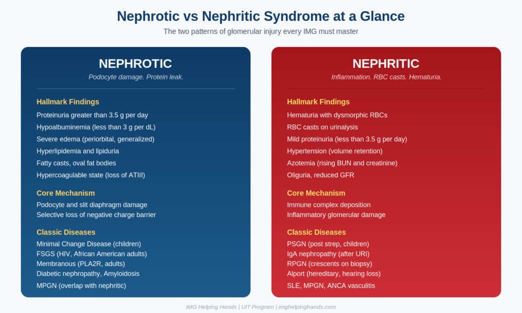
Figure 1. Nephrotic vs Nephritic syndromes at a glance. IMGHH UIT comparison.
| Feature | Nephrotic Syndrome | Nephritic Syndrome |
|---|---|---|
| Proteinuria | Massive, greater than 3.5 g per day | Mild to moderate, less than 3.5 g per day |
| Hematuria | Absent or microscopic | Present, often with RBC casts |
| Edema | Severe, periorbital, generalized | Mild, dependent |
| Hypertension | Usually absent or mild | Common and often significant |
| Hyperlipidemia and lipiduria | Classic finding | Absent |
| RBC casts | Absent | Hallmark feature |
| Oval fat bodies and fatty casts | Hallmark feature | Absent |
| GFR | Often preserved early | Reduced, leading to azotemia and oliguria |
| Complement (C3 and C4) | Usually normal | May be low (PSGN, SLE, MPGN, endocarditis) |
| Immune mechanism | Podocyte injury, charge barrier loss | Immune complex deposition, anti GBM, ANCA |
| Biopsy hallmark | Foot process effacement, deposits | Hypercellularity, crescents, necrosis |
| Hypercoagulability | Yes (loss of antithrombin III) | No |
| Infections risk | Increased (loss of IgG) | Not directly increased |
| Exam Pearl If you see massive proteinuria, edema, and high cholesterol in a vignette without hematuria, that is nephrotic. If you see cola colored urine, hypertension, and RBC casts, that is nephritic. The diseases follow the pattern. Find the pattern first, then pick the disease. |
To understand glomerular disease, you need to understand the glomerular filtration barrier. Every drop of blood that passes through the kidney is filtered through three layers stacked on top of each other.
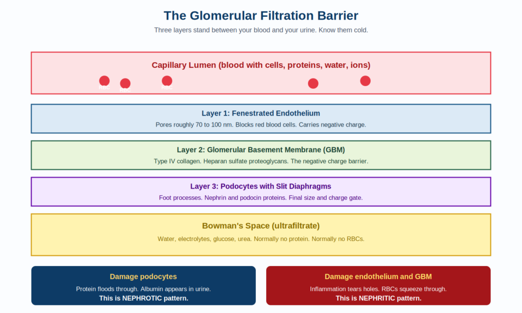
Figure 2. The three layered glomerular filtration barrier and what happens when each layer is damaged.
Imagine you are pouring water through three coffee filters stacked on top of each other. Each filter has a slightly smaller pore size and a different charge. Together they keep proteins and cells in the blood while letting water, salts, glucose, and urea pass through into the urine.
Damage to the podocytes (or the slit diaphragm proteins) opens the barrier to negatively charged albumin. Result: massive proteinuria, often selective for albumin in early disease. The endothelium and GBM remain intact, so red blood cells do not leak through. That is why nephrotic syndrome usually has no hematuria.
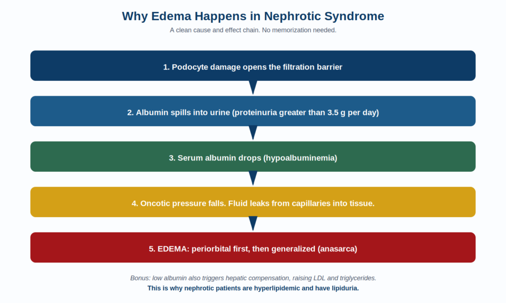
Figure 3. The cascade from proteinuria to edema. Memorize this sequence, not random facts.
Nephritic syndrome is fundamentally a problem of inflammation, not protein leak. Something triggers immune complex deposition or direct antibody binding in the glomerulus. Neutrophils, macrophages, and complement get recruited. The inflammatory infiltrate physically damages the endothelium and GBM. Red blood cells slip through the holes. As they pass through the distal tubule they get squeezed and dysmorphic, sometimes forming RBC casts.
At the same time, glomerular capillary loops are compressed and obstructed. GFR drops. Sodium and water are retained. Blood pressure rises. The patient becomes oliguric and azotemic.
| High Yield Trap Students often think hypertension in nephritic syndrome is from volume retention alone. There is also a renin angiotensin component because reduced GFR triggers the juxtaglomerular apparatus. NBME questions sometimes test this through plasma renin activity. |
Urinalysis is the single most cost effective tool in nephrology. A good IMG can solve half a renal vignette before getting to the biopsy. Here is how to read it.
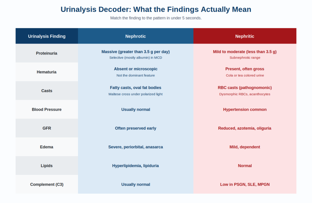
Figure 4. How to translate urinalysis findings into the correct syndrome bucket.
A cast is a cylindrical structure formed in the renal tubule. The matrix is Tamm Horsfall protein. Whatever gets trapped in that matrix gives the cast its identity. Each cast type points to a specific disease pattern.
| Cast Type | What It Means |
|---|---|
| RBC casts | Glomerulonephritis (nephritic). Pathognomonic. |
| WBC casts | Pyelonephritis or acute interstitial nephritis. |
| Fatty casts and oval fat bodies | Nephrotic syndrome. Maltese cross appearance under polarized light. |
| Muddy brown granular casts | Acute tubular necrosis. |
| Waxy casts | Chronic kidney disease, end stage renal disease. |
| Hyaline casts | Benign. Seen with dehydration, exercise, fever. |
Five diseases account for the vast majority of nephrotic vignettes on Step 1. We will cover each one with the same scaffolding: who gets it, what triggers it, how it looks under each microscope, classic vignette clues, and the NBME traps to watch for.
The most common cause of nephrotic syndrome in children, usually between ages 2 and 8.
Reversible podocyte foot process effacement, likely T cell mediated. Cytokines damage the slit diaphragm but the glomerulus looks completely normal on light microscopy. Hence the name.
A 5 year old boy presents with periorbital edema that started 2 weeks after an upper respiratory infection. He has been gaining weight and his clothes feel tight. Urinalysis shows 4+ protein, no blood, no casts beyond fatty ones. Serum albumin is 2.1 g per dL. Cholesterol is 380 mg per dL.
Excellent response to corticosteroids. Most children remit fully within 4 to 6 weeks. Some relapse. Steroid resistant cases may turn out to be FSGS in disguise.
| NBME Trap for MCD Selective albuminuria is highly suggestive of MCD because the charge barrier is the main thing damaged. You lose albumin (small, negatively charged) but retain larger proteins. If the question shows selectivity, lean MCD. |
The most common cause of primary nephrotic syndrome in adults in the United States, especially among African American patients.
Podocyte injury leading to segmental sclerosis in some (not all) glomeruli. Primary FSGS is idiopathic and may involve a circulating permeability factor. Secondary FSGS is associated with HIV (collapsing variant), heroin use, obesity, sickle cell disease, and reflux nephropathy.
A 32 year old African American man with HIV not on antiretrovirals presents with bilateral lower extremity edema and 8 g per day proteinuria. He is mildly hypertensive. Biopsy shows segmental sclerosis in a portion of the glomeruli.
Less responsive to steroids than MCD. Progresses to ESRD in many patients. ACE inhibitors and ARBs are core therapy.
The most common cause of primary nephrotic syndrome in white adults.
Antibodies (most commonly against the PLA2R antigen on podocytes in primary disease) form subepithelial immune complexes. The complexes are visible as electron dense deposits between the GBM and podocyte. New GBM grows around them, creating the classic spike and dome appearance.
A 55 year old man presents with new lower extremity edema and 5 g per day proteinuria. He has had unexplained weight loss. PLA2R antibody is positive. Biopsy shows diffuse thickening of capillary loops with spike and dome appearance on silver stain.
Membranous nephropathy has the highest risk of renal vein thrombosis among nephrotic syndromes because of profound antithrombin III loss. Sudden flank pain with hematuria is a board favorite presentation.
The most common cause of chronic kidney disease and end stage renal disease in the United States overall.
Chronic hyperglycemia causes non enzymatic glycosylation of the GBM. Mesangial matrix expands. Eventually nodular sclerosis develops (Kimmelstiel Wilson nodules). The earliest finding is hyperfiltration, then microalbuminuria, then frank proteinuria, then declining GFR.
A 58 year old man with poorly controlled type 2 diabetes for 20 years presents with 6 g per day proteinuria and mild edema. He has retinopathy and stocking glove neuropathy. Biopsy would show nodular glomerulosclerosis.
ACE inhibitors and ARBs slow progression by reducing intraglomerular pressure. SGLT2 inhibitors have transformed the prognosis over the last decade and are now first line for diabetic kidney disease.
Deposition of misfolded protein in the mesangium and along capillary walls. AL amyloid (light chains in multiple myeloma) is the most common in the United States. AA amyloid (serum amyloid A) is associated with chronic inflammatory diseases like rheumatoid arthritis and familial Mediterranean fever.
A 67 year old man with multiple myeloma develops new edema and 10 g per day proteinuria. Biopsy shows apple green birefringence under polarized light after Congo red staining.
| Quick Recognition Pattern for Nephrotic Diseases 1. Child plus edema after URI: Minimal Change Disease. 2. African American or HIV adult: FSGS. 3. Adult, PLA2R positive, hepatitis B, or solid tumor: Membranous. 4. Long standing diabetes, retinopathy, neuropathy: Diabetic nephropathy. 5. Multiple myeloma or chronic inflammation: Amyloidosis. |
Five nephritic syndromes dominate Step 1. The same teaching scaffold applies: who, why, how it presents, what biopsy shows, and the NBME traps.
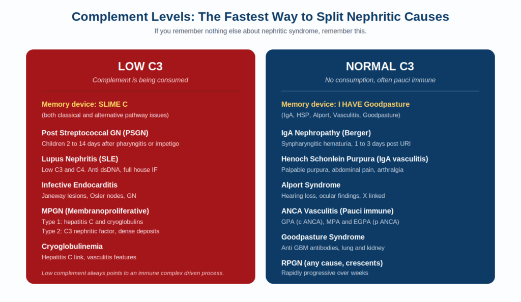
Figure 5. The single most useful nephritic decision split: complement levels.
Before diving into individual diseases, lock in the complement split above. C3 low versus C3 normal cuts the nephritic differential in half before you read a single biopsy result.
The classic pediatric nephritic syndrome. Self limiting in most children.
Immune complexes containing nephritogenic strains of group A beta hemolytic Streptococcus (often pyrogenic exotoxin B) deposit in the subepithelial space. Complement is activated and consumed. The result is acute, inflammatory damage with classic subepithelial humps on electron microscopy.
A 7 year old boy presents with cola colored urine, periorbital edema, and elevated blood pressure 2 weeks after a sore throat. Urinalysis shows RBC casts and proteinuria. Anti DNase B and ASO titers are elevated. C3 is low.
Children recover fully in over 95 percent of cases. Adults have a more guarded outcome and may progress to chronic kidney disease.
The most common primary glomerulonephritis worldwide.
Abnormally glycosylated IgA1 deposits in the mesangium. Triggers mesangial inflammation. The mucosal immune system overproduces IgA after upper respiratory or gastrointestinal infection, which is why episodes coincide with infection (synpharyngitic).
A 24 year old man presents with gross hematuria 1 to 3 days after the onset of an upper respiratory infection. Episodes have happened twice before. C3 is normal. Biopsy shows IgA deposits in the mesangium.
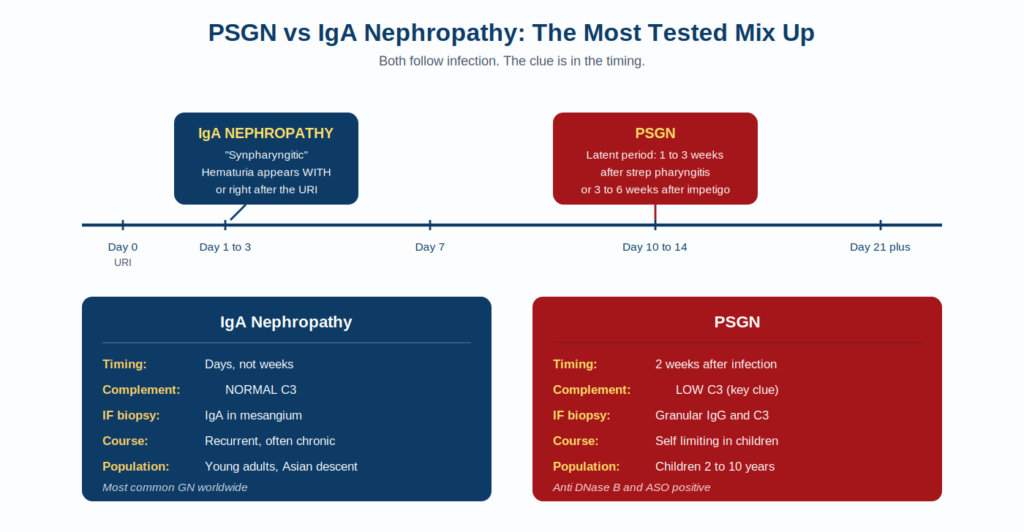
Figure 6. The single most important Step 1 mix up: PSGN vs IgA timing.
Variable. Up to 30 percent progress to ESRD over decades.
Hereditary defect (most commonly X linked) in type IV collagen, specifically the alpha 5 chain (COL4A5). The GBM cannot form properly. Result: a thinned, irregular, split, basket weave appearance on electron microscopy.
A 14 year old boy presents with persistent microscopic hematuria. He has sensorineural hearing loss and a family history of kidney failure. Eye examination shows anterior lenticonus.
Not one disease. A clinical pattern of rapid decline in renal function over weeks, with crescents on biopsy. Three immune mechanisms cause it.
| Pattern | Immunofluorescence | Examples |
|---|---|---|
| Anti GBM antibody | Linear IgG | Goodpasture syndrome (lung and kidney) |
| Immune complex | Granular IgG and C3 | PSGN, SLE, IgA, HSP |
| Pauci immune | Negative or minimal | ANCA vasculitis: GPA, MPA, EGPA |
Anti GBM antibodies cross react with alveolar basement membrane. Result: hemoptysis plus rapidly progressive renal failure in a young male smoker.
Upper airway (sinusitis, nasal septal perforation, saddle nose), lung (cavitary lesions), and kidney involvement. c ANCA positive. Treat with steroids plus cyclophosphamide or rituximab.
p ANCA positive. EGPA also has asthma and eosinophilia.
MPGN is a glomerular pattern that can cause both nephrotic and nephritic features. It is a hybrid syndrome.
Tram track appearance on silver stain (double contour of the GBM from mesangial interposition).
| Nephritic Quick Recognition 1. Child 2 weeks after sore throat, cola urine, low C3: PSGN. 2. Young adult with gross hematuria DURING a URI, normal C3: IgA nephropathy. 3. Boy with hematuria, deafness, and a positive family history: Alport syndrome. 4. Hemoptysis plus renal failure, linear IF: Goodpasture. 5. Sinusitis, lung cavities, kidney, c ANCA: GPA. 6. Hepatitis C, cryoglobulins, tram track: MPGN type 1. |
This section is what separates 250 scorers from 270 scorers on Step 1 renal questions. Memorize this matrix. Every glomerular disease has a signature on light microscopy (LM), immunofluorescence (IF), and electron microscopy (EM). Once you know the signature, the diagnosis is automatic.
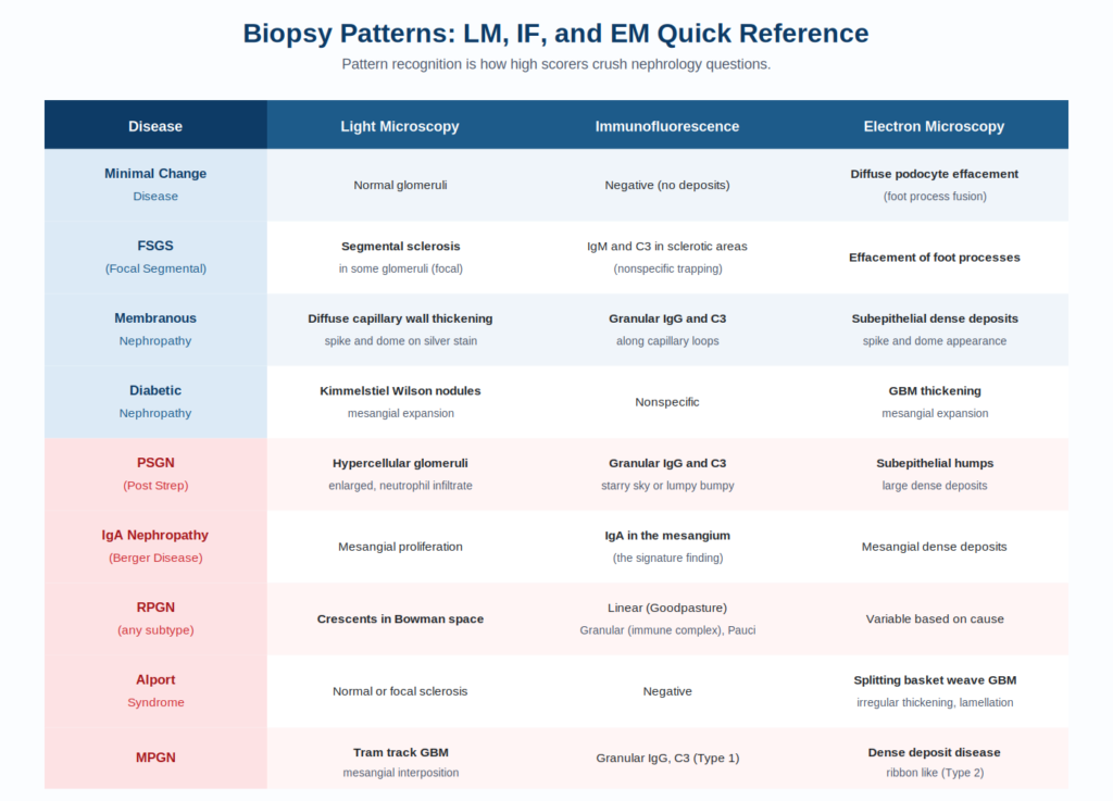
Figure 7. Biopsy pattern matrix for all major glomerular diseases.
Mnemonics work when they are vivid, when they map cleanly to the content, and when you build them yourself. The ones below have been refined across hundreds of UIT tutoring sessions. They stick.
Five nephrotic diseases. One acronym. Five seconds to recall.
M Minimal Change Disease (children, steroid responsive)
F FSGS (HIV, African American, heroin)
M Membranous (PLA2R, hepatitis B, solid tumors)
D Diabetic nephropathy (Kimmelstiel Wilson)
A Amyloidosis (Congo red, apple green birefringence)
M MPGN (overlap, tram track, dense deposit)
Whenever the complement is low, run through this list.
S SLE (systemic lupus, full house pattern, anti dsDNA)
L Lupus is often listed separately for emphasis
I Infective endocarditis (Janeway, Osler, GN)
M MPGN (especially type 2, dense deposit disease)
E Endocarditis or Endo cryoglobulinemia
C Cryoglobulinemia (hepatitis C link)
Plus PSGN (post streptococcal)
Whenever complement is normal in a nephritic vignette, run through this list.
I IgA nephropathy (synpharyngitic hematuria)
H Henoch Schonlein Purpura (palpable purpura, IgA vasculitis)
A Alport syndrome (hearing loss, basket weave)
V Vasculitis with ANCA (GPA, MPA, EGPA)
E (any vasculitis with normal complement)
G Goodpasture syndrome (anti GBM, hemoptysis)
| Buzzword | Diagnosis |
|---|---|
| Spike and dome (silver stain) | Membranous nephropathy |
| Subepithelial humps | PSGN |
| Tram track | MPGN |
| Basket weave or splitting GBM | Alport syndrome |
| Linear IgG along GBM | Goodpasture syndrome |
| Lumpy bumpy or starry sky | PSGN |
| Mesangial IgA deposits | IgA nephropathy |
| Crescents in Bowman space | RPGN |
| Foot process effacement (normal LM) | Minimal Change Disease |
| Kimmelstiel Wilson nodules | Diabetic nephropathy |
| Apple green birefringence | Amyloidosis |
| Wire loop appearance | Lupus nephritis (Class IV) |
Over the years, NBME questions on glomerular disease have followed a small number of repeatable templates. Recognize the template and you save 30 seconds per question.
Vignette gives you classic findings (proteinuria 5 g, edema, hyperlipidemia OR cola urine, hypertension, RBC casts). Asks you to pick the most likely diagnosis. Just match the pattern to the most common cause in that demographic.
Vignette is short. The image or description of the biopsy is the actual question. EM findings dominate here. Subepithelial humps, basket weave, foot process effacement, spike and dome, dense deposits are the high yield words.
Vignette includes a serum complement level. If low, narrow to the SLIME C list. If normal, narrow to the I HAVE Goodpasture list. The complement level is doing all the heavy lifting.
Vignette describes nephrotic findings, then asks about a complication. Renal vein thrombosis (membranous), infection with pneumococcus (loss of IgG), hyperlipidemia, hypocalcemia (loss of vitamin D binding protein). Each complication maps back to a lost protein.
Asks you why something happens. Why edema? Falling oncotic pressure. Why hyperlipidemia? Liver compensation for albumin loss. Why hypercoagulability? Antithrombin III loss. Always link back to the original protein leak.
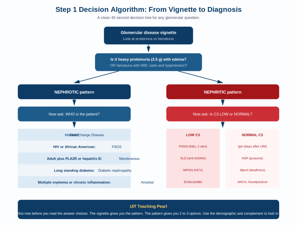
Figure 8. The decision algorithm we recommend using on every glomerular Step 1 question.
Most prep resources give you content. We give you a system. The Ultimate Integrated Teaching (UIT) approach at IMG Helping Hands is built around four principles that change how you absorb medicine.
If you can derive a fact from a mechanism, you do not need to memorize it. Edema in nephrotic syndrome is not a fact. It is a consequence of falling oncotic pressure. Hyperlipidemia is not a fact. It is hepatic compensation. The fewer facts you memorize, the more you can recall under pressure.
Nephrology cannot be studied in isolation. PSGN connects to microbiology (group A strep). Goodpasture connects to immunology (autoantibodies). Amyloid connects to hematology (multiple myeloma). Our teaching explicitly draws these threads so that one renal question reinforces three other topics.
Every UIT lesson is built around a small set of master diagrams. The filtration barrier diagram. The complement split. The biopsy matrix. When you sit down for Step 1, your brain pulls up an image, not a sentence. Pattern recognition beats verbal recall every time.
We do not just teach the material. We teach the question. NBME vignettes follow rules. The first sentence tells you the age and sex. The second sentence usually buries the trigger. The lab values are designed to push you toward or away from a diagnosis. Knowing these patterns is half the battle.
| How to Learn Like a UIT Student 1. Read each section here once for understanding. 2. Re read the comparison table and the biopsy matrix daily for one week. 3. Take the practice MCQs at the end of this guide without notes. 4. Whatever you get wrong, return to that disease’s section and rebuild the mechanism. 5. Teach the topic to a friend out loud. If you can teach it, you own it. |
Read this section the night before. It is everything you need in one page.
Fifteen original questions. No looking at your notes. Time yourself: 90 seconds per question. Mark the ones you miss and revisit those sections.
Question 1. A 5 year old boy is brought to the pediatrician because of swelling around the eyes that has been worsening for 1 week. His mother reports he had an upper respiratory infection 2 weeks ago. His blood pressure is normal. Urinalysis shows 4+ protein and fatty casts. Serum albumin is 2.0 g per dL. Serum cholesterol is 410 mg per dL. Renal biopsy shows normal glomeruli on light microscopy and electron microscopy shows diffuse foot process effacement. Which of the following is the most appropriate initial treatment?
(A) Cyclophosphamide
(B) Lisinopril
(C) Plasmapheresis
(D) Prednisone
(E) Rituximab
Answer: D. Prednisone
Explanation: This is classic minimal change disease (MCD). Pediatric age, abrupt nephrotic syndrome after a URI, normal light microscopy, foot process effacement on EM. MCD is exquisitely steroid responsive and corticosteroids are first line. Cyclophosphamide and rituximab are reserved for frequent relapsers or steroid resistant cases. Plasmapheresis is used in Goodpasture and TTP, not MCD. Lisinopril would be used in chronic proteinuric kidney disease but is not the initial treatment for MCD.
Question 2. A 28 year old man presents with gross hematuria. He reports the urine became dark red 1 day after the start of an upper respiratory infection. He had a similar episode 18 months ago, also during a cold. Blood pressure is 132 over 84. Urinalysis shows 3+ blood, 1+ protein, and dysmorphic red blood cells. Serum creatinine is 1.2 mg per dL. Serum C3 and C4 are normal. Which of the following findings would most likely be present on renal biopsy?
(A) Anti glomerular basement membrane antibodies (linear IgG)
(B) Crescents in Bowman space
(C) IgA deposition in the mesangium
(D) Subepithelial electron dense humps
(E) Wire loop appearance on light microscopy
Answer: C. IgA deposition in the mesangium
Explanation: Synpharyngitic hematuria (hematuria coinciding with or appearing 1 to 3 days after a URI), normal complement, and recurrent episodes in a young adult are the textbook triad for IgA nephropathy. Mesangial IgA deposits are the hallmark on immunofluorescence. Subepithelial humps would suggest PSGN, which presents 2 weeks (not 1 day) after pharyngitis and has LOW C3. Linear IgG suggests Goodpasture. Crescents indicate RPGN. Wire loop is lupus nephritis.
Question 3. A 7 year old girl presents 14 days after a sore throat with cola colored urine and periorbital edema. Blood pressure is 142 over 92. Urinalysis shows red blood cell casts and dysmorphic erythrocytes. Serum creatinine is 1.6 mg per dL. Anti DNase B titers are elevated. Serum C3 is markedly decreased. Which of the following best describes the expected electron microscopy finding?
(A) Basket weave splitting of the glomerular basement membrane
(B) Diffuse foot process effacement only
(C) Mesangial dense deposits
(D) Subendothelial deposits with mesangial interposition
(E) Subepithelial electron dense humps
Answer: E. Subepithelial electron dense humps
Explanation: Classic PSGN. Pediatric patient, 2 week latent period after streptococcal infection, hematuria with RBC casts, low C3, elevated streptococcal antibodies. EM shows large subepithelial dense deposits (humps). Basket weave indicates Alport. Foot process effacement alone is MCD. Mesangial deposits suggest IgA. Subendothelial deposits with tram track point to MPGN.
Question 4. A 56 year old man presents with new onset lower extremity edema and 6 g per day of proteinuria. He has lost 5 kg unintentionally over the past 4 months. PLA2R antibody is positive. Renal biopsy on silver stain shows diffuse capillary loop thickening with a spike and dome appearance. Which of the following is the most likely associated complication?
(A) Bone marrow failure
(B) Diabetic ketoacidosis
(C) Hepatic encephalopathy
(D) Pulmonary hemorrhage
(E) Renal vein thrombosis
Answer: E. Renal vein thrombosis
Explanation: Classic membranous nephropathy. The patient should also be evaluated for occult malignancy given his age and weight loss. Membranous nephropathy carries the highest risk of renal vein thrombosis among nephrotic syndromes because of marked antithrombin III loss. Watch for sudden flank pain with gross hematuria. Pulmonary hemorrhage suggests Goodpasture, not membranous.
Question 5. A 32 year old African American man with HIV (not on antiretroviral therapy) presents with bilateral lower extremity edema and 9 g per day of proteinuria. Serum creatinine is 1.8 mg per dL. Renal biopsy shows segmental sclerosis in 30 percent of the glomeruli. Which of the following is the most likely diagnosis?
(A) Diabetic nephropathy
(B) Focal segmental glomerulosclerosis
(C) IgA nephropathy
(D) Membranous nephropathy
(E) Minimal change disease
Answer: B. Focal segmental glomerulosclerosis
Explanation: HIV positive plus African American plus heavy proteinuria plus segmental (focal) sclerosis on biopsy points directly to FSGS, often the collapsing variant in HIV. FSGS is the most common cause of primary nephrotic syndrome in African American adults. The biopsy description is pathognomonic.
Question 6. A 14 year old boy is referred for evaluation of microscopic hematuria found on a school physical. He has bilateral sensorineural hearing loss. His maternal uncle died of kidney failure at age 35. Slit lamp examination shows anterior lenticonus. Which of the following defects is most likely responsible for his condition?
(A) Anti glomerular basement membrane antibodies
(B) Defective complement regulation
(C) IgA glycosylation abnormality
(D) Mutation in type IV collagen
(E) Podocyte slit diaphragm protein defect
Answer: D. Mutation in type IV collagen
Explanation: Alport syndrome. Triad of hematuria, sensorineural deafness, and ocular abnormalities (anterior lenticonus). X linked inheritance is most common (defect in COL4A5). EM shows basket weave splitting of the GBM. Podocyte slit diaphragm defects cause congenital nephrotic syndrome of the Finnish type (nephrin mutation).
Question 7. A 22 year old male smoker presents with hemoptysis and rapidly worsening renal function. Serum creatinine is 5.2 mg per dL (up from 1.0 two months ago). Urinalysis shows RBC casts. Chest imaging shows bilateral patchy infiltrates. Anti GBM antibody is positive. Which of the following is the most likely immunofluorescence pattern on renal biopsy?
(A) Granular IgG and C3 along capillary loops
(B) IgA in the mesangium
(C) Linear IgG along the glomerular basement membrane
(D) Pauci immune (negative) staining
(E) Subepithelial humps
Answer: C. Linear IgG along the glomerular basement membrane
Explanation: Goodpasture syndrome. Anti GBM antibodies bind alpha 3 chain of type IV collagen in both alveolar and glomerular basement membranes, producing pulmonary renal syndrome. The IF pattern is linear (smooth, ribbon like), unlike the granular pattern of immune complex disease. Smoking is a known risk factor.
Question 8. A 45 year old woman with chronic hepatitis C presents with new onset edema, hypertension, and 4 g per day of proteinuria. Serum creatinine is 2.1 mg per dL. Serum C3 and C4 are decreased. Cryoglobulins are positive. Which of the following findings would be expected on renal biopsy?
(A) Apple green birefringence with Congo red staining
(B) Basket weave splitting of the GBM
(C) Foot process effacement without immune deposits
(D) Kimmelstiel Wilson nodules
(E) Tram track appearance on silver stain
Answer: E. Tram track appearance on silver stain
Explanation: MPGN type 1, classically associated with hepatitis C and cryoglobulinemia. Mesangial interposition between the endothelial cell and the GBM creates a double contour (tram track) on silver stain. Apple green birefringence is amyloidosis. Basket weave is Alport. Foot process effacement only is MCD. Kimmelstiel Wilson is diabetic nephropathy.
Question 9. A 58 year old man with a 20 year history of type 2 diabetes is referred for evaluation of progressive proteinuria. His urine albumin to creatinine ratio is 2800 mg per g. He has diabetic retinopathy and peripheral neuropathy. Which of the following pathologic findings is most likely present in his kidney?
(A) Crescents in Bowman space
(B) Linear IgG along the GBM
(C) Mesangial IgA deposits
(D) Nodular glomerulosclerosis (Kimmelstiel Wilson)
(E) Subepithelial humps
Answer: D. Nodular glomerulosclerosis (Kimmelstiel Wilson)
Explanation: Long standing diabetes with retinopathy and neuropathy strongly suggests diabetic nephropathy. The pathognomonic finding is nodular sclerosis (Kimmelstiel Wilson nodules) on light microscopy. EM shows GBM thickening and mesangial expansion.
Question 10. A 65 year old man with multiple myeloma develops new lower extremity edema and 11 g per day of proteinuria. Renal biopsy with Congo red staining shows apple green birefringence under polarized light. Which of the following is the most likely composition of the deposited material?
(A) Amyloid A protein
(B) Beta 2 microglobulin
(C) Immunoglobulin light chains
(D) Mutant transthyretin
(E) Prion protein
Answer: C. Immunoglobulin light chains
Explanation: Renal amyloidosis with apple green birefringence under polarized light is the hallmark. In multiple myeloma, the deposited amyloid is composed of immunoglobulin light chains (AL amyloid). Amyloid A (AA) is from chronic inflammation. Beta 2 microglobulin amyloid occurs in long term dialysis.
Question 11. A 28 year old woman with systemic lupus erythematosus presents with new onset hypertension, edema, and 5 g per day of proteinuria. Urinalysis shows RBC casts. Serum C3 and C4 are decreased. Anti dsDNA antibodies are strongly positive. Renal biopsy is most likely to show which of the following immunofluorescence patterns?
(A) Anti GBM linear IgG
(B) Full house staining (IgG, IgA, IgM, C3, C1q)
(C) IgA deposits in the mesangium only
(D) No immune deposits (pauci immune)
(E) Subepithelial humps only
Answer: B. Full house staining (IgG, IgA, IgM, C3, C1q)
Explanation: Lupus nephritis classically shows full house immunofluorescence reflecting deposition of all major immunoglobulin classes and complement components. Class IV (diffuse proliferative) lupus nephritis is the most common and most severe form, often presenting with both nephrotic and nephritic features. Low complement and positive anti dsDNA are the giveaways.
Question 12. A 62 year old man with chronic sinusitis, a saddle nose deformity, and cavitary lung lesions on chest imaging develops rapidly worsening renal function. Serum creatinine has risen from 1.1 to 4.8 mg per dL over 6 weeks. Urinalysis shows RBC casts. Serum C3 is normal. Which of the following serologic findings would most likely be positive?
(A) Anti GBM antibodies
(B) Anti dsDNA antibodies
(C) Anti PLA2R antibodies
(D) c ANCA (anti proteinase 3)
(E) Cryoglobulins
Answer: D. c ANCA (anti proteinase 3)
Explanation: Classic granulomatosis with polyangiitis (GPA, formerly Wegener). Upper airway (sinusitis, saddle nose), lung (cavities), and kidney (RPGN) involvement with c ANCA positivity (anti PR3) is the diagnostic constellation. RPGN here is pauci immune on biopsy.
Question 13. A 6 year old boy presents with palpable purpura on the lower extremities and buttocks, abdominal pain, arthralgias, and microscopic hematuria 1 week after an upper respiratory infection. Urinalysis shows RBC casts and mild proteinuria. Serum C3 is normal. Which of the following findings would most likely be present on renal biopsy?
(A) Anti GBM antibodies
(B) Crescents with pauci immune staining
(C) IgA deposits in the mesangium
(D) Linear IgG staining
(E) Subepithelial humps
Answer: C. IgA deposits in the mesangium
Explanation: Henoch Schonlein purpura (also called IgA vasculitis) is the systemic counterpart of IgA nephropathy. Same IgA deposits in the mesangium, same normal complement. Distinguishing features are palpable purpura, abdominal pain (intussusception risk), and arthralgia. Most common in children.
Question 14. A 38 year old man with nephrotic syndrome from membranous nephropathy presents with sudden onset left flank pain and gross hematuria. Renal ultrasound with Doppler shows reduced flow in the left renal vein. Which of the following best explains this complication?
(A) Direct injury from glomerular inflammation
(B) Loss of antithrombin III in the urine
(C) Renal artery atherosclerosis
(D) Severe hypertension causing endothelial damage
(E) Vasculitis affecting the renal vasculature
Answer: B. Loss of antithrombin III in the urine
Explanation: Membranous nephropathy carries the highest risk of renal vein thrombosis among nephrotic syndromes. Mechanism: urinary loss of antithrombin III creates a hypercoagulable state. Patients also lose plasminogen and gain procoagulants from hepatic compensation. Presentation: sudden flank pain plus hematuria plus worsening proteinuria.
Question 15. A 30 year old man presents 12 days after a sore throat with cola colored urine, blood pressure 158 over 96, and periorbital edema. Urinalysis shows RBC casts. Serum complement studies show: C3 markedly decreased, C4 normal. Which of the following best explains the complement profile?
(A) Activation of the alternative pathway
(B) Activation of the lectin pathway
(C) Defective complement regulation
(D) Hereditary C3 deficiency
(E) Increased complement synthesis
Answer: A. Activation of the alternative pathway
Explanation: PSGN consumes complement primarily through the alternative pathway, which uses C3 but not C4. Low C3 with normal C4 is characteristic. In contrast, SLE activates the classical pathway, consuming both C3 and C4. Recognizing the C3 low, C4 normal pattern is a high yield Step 1 distinction.
These are the questions IMG students ask us most often during UIT tutoring sessions.
Albumin leaks into urine. Serum albumin drops. Plasma oncotic pressure falls. Fluid moves from the capillaries into the interstitial space following Starling forces. The result is edema, classically periorbital first (because facial tissue is loose), then generalized (anasarca). A secondary mechanism involves primary renal sodium and water retention.
Inflammation creates holes in the glomerular filtration barrier. Red blood cells squeeze through into Bowman space. As they travel through the tubule, they become trapped in Tamm Horsfall protein and get molded into a cylindrical cast. The combination of RBCs plus the cylindrical Tamm Horsfall matrix is what gives you the classic RBC cast on urinalysis. RBC casts are pathognomonic for glomerular bleeding.
If we had to pick just one, it would be Minimal Change Disease, because it is the most commonly tested in pediatric vignettes and the EM finding (foot process effacement) is a frequent answer choice. FSGS comes a close second, particularly in HIV positive or African American adult vignettes. Membranous nephropathy is also high yield, especially the renal vein thrombosis complication.
PSGN and IgA nephropathy are tied for first place. PSGN tests pattern recognition (child, 2 weeks after sore throat, low C3) and complement biology. IgA nephropathy tests timing (synpharyngitic vs latent) and immunofluorescence (mesangial IgA). After those two, RPGN with anti GBM disease (Goodpasture) and ANCA vasculitis are next in frequency.
Nephrotic loses protein because podocytes are damaged. Nephritic loses blood because inflammation tears the filter. Protein loss leads to edema, hyperlipidemia, and hypercoagulability. Blood loss leads to hematuria, RBC casts, hypertension, and azotemia. That single sentence will get you through 90 percent of Step 1 glomerular questions.
Timing and complement. PSGN comes 2 weeks after pharyngitis or 3 to 6 weeks after impetigo, with low C3. IgA comes 1 to 3 days after a URI (synpharyngitic), with normal C3. Age also helps: PSGN is mostly children, IgA is mostly young adults. Biopsy is the definitive answer: IgA shows mesangial IgA deposits, PSGN shows subepithelial humps.
Three diseases are notorious for crossing the line: MPGN, lupus nephritis (especially Class IV), and diffuse proliferative GN. When you see mixed features, lean toward one of these. MPGN gives you tram track and low complement. Lupus gives you full house IF and multi system disease. Both are high yield.
Loop diuretics are commonly used to control edema but must be used carefully because patients are intravascularly volume depleted despite massive total body fluid overload. Aggressive diuresis can cause prerenal injury. ACE inhibitors and ARBs are core therapy to reduce proteinuria. Statins manage hyperlipidemia. Anticoagulation is considered when serum albumin drops below 2 g per dL, especially in membranous nephropathy.
Hepatitis B antigens form subepithelial immune complexes that deposit in the GBM, producing membranous nephropathy. Hepatitis C is associated with cryoglobulins (immune complexes that precipitate in the cold), which deposit subendothelially and trigger MPGN type 1. Memorize the pairing: B for membranous, C for MPGN.
Pauci means few. Pauci immune means immunofluorescence shows few or no immune deposits, even though crescents are forming. The classic example is ANCA associated vasculitis (GPA, MPA, EGPA). It tells you the damage is neutrophil mediated rather than immune complex mediated.
If you have read this guide from start to finish, you now understand something that most IMG students never quite grasp before their exam. Nephrology is not a memorization game. It is a pattern recognition game played on top of a small number of clean mechanisms. Once you have those mechanisms locked in, the buzzwords, biopsy findings, and answer choices all start to look obvious.
Here is what we want you to take away:
At IMG Helping Hands, we believe medical education for IMGs deserves more than fragmented PDFs and recycled study notes. The UIT (Ultimate Integrated Teaching) program is built on the conviction that you learn faster when teaching is integrated, visual, mechanism first, and exam aligned. We have seen students go from struggling with renal pathology to scoring in the top deciles using exactly the framework you have just read.
If this guide helped you, share it with the IMG who needs it most. Save it for the night before your exam. Come back to it after rotations when you need a quick refresh. And know that on the other side of this exam is a residency program that needs exactly the kind of clinician this study process is shaping you to become.
CONTINUE YOUR USMLE JOURNEY WITH IMG HELPING HANDS
Explore the UIT program and structured USMLE Step 1 review system at IMG Helping Hands, designed specifically for international medical graduates preparing for long-term exam success.
Access companion study plan guides for 3-month, 6-month, and 12-month Step 1 preparation, along with high-yield organ system reviews covering cardiology, pulmonology, GI, endocrine, hematology, and more using the same UIT framework.
Subscribe for new weekly educational content built specifically for IMGs.
Structured learning. Integrated systems. Built for IMG success.
This guide is built from a synthesis of current nephrology and pathology evidence. For deeper reading we recommend:
Articles published by IMG Helping Hands are prepared by our team using information from direct experience, publicly available resources, and educational references. AI tools may be used to assist with drafting, proofreading, and formatting; however, all content undergoes review and approval before publication.
The information provided is intended for educational purposes only. Requirements, policies, and processes may change over time. Readers should consult official sources for the most current information.