

Rate limiting enzymes feel chaotic for one reason. Most of us learned each pathway in a different week, from a different lecturer, and never sat down to line the regulators up side by side. When you do that, the chaos disappears. There are fifteen enzymes. The regulators repeat. Two hormones and one chemical trick explain almost every question. This is the same pattern-first approach that makes other dense enzyme topics like the lysosomal storage diseases click, learn the one rule, then every vignette that dresses it up. This guide is that lineup, and nothing else.
We rebuilt this from a much longer draft with one filter: does this line change an answer on Step 1? What survived is the fifteen enzyme master list, the fed versus fasting framework, the one molecule that flips glycolysis and gluconeogenesis at the same time, the drugs and toxins that act at these enzymes, and the diseases the exam attaches to them. What we cut is the full mechanism of each reaction, the minor allosteric effectors that never appear in a stem, the cross talk between AMPK and every pathway it touches, and the historical detail on how each enzyme was characterized. If you want the biochemistry textbook version, it exists. This is the exam version.
A rate limiting enzyme is the slowest step in a pathway, so the whole pathway can only run as fast as that step. It is usually the first irreversible reaction, the point after which the substrate is committed. And because controlling this one step controls everything downstream, it is where the cell puts its regulation. Allosteric effectors, hormonal phosphorylation, and drug targets all converge here. That is why the exam asks about it. One enzyme tells you the metabolic state, the regulators, the drug, and often the disease.
Two kinds of regulation matter. Allosteric control is fast and local: ATP, AMP, citrate, acetyl CoA and the pathway’s own end product bind the enzyme and shift its activity within seconds. Hormonal control is slower but sets the whole body’s direction. Insulin and glucagon act through phosphorylation cascades, and the rule that resolves most fed versus fasting questions is this: phosphorylation switches on breakdown enzymes and switches off synthesis enzymes. Glucagon adds phosphate, insulin removes it.
| How We Teach This Before students memorize a single regulator, we make them answer one question about every vignette: is this patient fed or fasting? Fed means insulin, dephosphorylation, storage. Fasting means glucagon, phosphorylation, breakdown. Once that is settled, the enzyme falls out. Most students who miss these questions never asked the question. They went straight to the answer choices and picked the enzyme that sounded most important. |
Figure 1 puts all fifteen enzymes on one page, colored by the state that activates them. Teal enzymes run when insulin is high. Orange enzymes run when glucagon is high. Navy enzymes are regulated by something other than the fed and fasting switch.
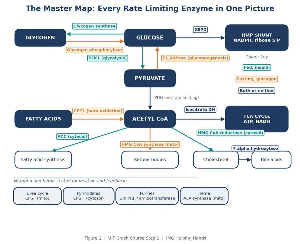
Figure 1. Every rate limiting enzyme, placed where it acts and colored by the hormonal state that turns it on.
Table 1 is the master list. Every question in this category can be answered from it, so it is worth rebuilding from memory on the whiteboard the morning of the exam.
| Pathway | Rate limiting enzyme | Turned up by | Turned down by | Hormone |
|---|---|---|---|---|
| Glycolysis | PFK1 | AMP, F2,6BP | ATP, citrate | Insulin up, glucagon down |
| Gluconeogenesis | Fructose 1,6 bisphosphatase | ATP, acetyl CoA, citrate | AMP, F2,6BP | Glucagon up, insulin down |
| TCA cycle | Isocitrate dehydrogenase | ADP, calcium | ATP, NADH | Energy state, not hormones |
| Glycogenesis | Glycogen synthase | G6P, insulin (dephosphorylated) | Glucagon, epinephrine (phosphorylated) | Insulin up, glucagon down |
| Glycogenolysis | Glycogen phosphorylase | AMP, glucagon, epinephrine (phosphorylated) | ATP, G6P, insulin | Glucagon up, insulin down |
| HMP shunt | Glucose 6 phosphate dehydrogenase | NADP+, insulin | NADPH | Insulin up |
| Pyrimidine synthesis | Carbamoyl phosphate synthetase II (cytosol) | ATP, PRPP | UTP | None tested |
| Purine synthesis | Glutamine PRPP amidotransferase | PRPP | AMP, GMP, IMP | None tested |
| Urea cycle | Carbamoyl phosphate synthetase I (mitochondria) | N acetylglutamate | None tested | None tested |
| Fatty acid synthesis | Acetyl CoA carboxylase | Citrate, insulin (dephosphorylated) | Palmitoyl CoA, glucagon (phosphorylated) | Insulin up, glucagon down |
| Fatty acid oxidation | Carnitine palmitoyltransferase I (CPT1) | Glucagon (indirectly) | Malonyl CoA | Glucagon up, insulin down |
| Ketogenesis | HMG CoA synthase (mitochondrial) | Glucagon, fatty acids | Insulin | Glucagon up, insulin down |
| Cholesterol synthesis | HMG CoA reductase | Insulin (dephosphorylated) | Glucagon, cholesterol, statins | Insulin up, glucagon down |
| Heme synthesis | ALA synthase (mitochondrial) | Drugs that consume heme | Heme, glucose | None tested |
| Bile acid synthesis | Cholesterol 7 alpha hydroxylase | Cholesterol | Bile acids | None tested |
USMLE Impact Theory teaches every high-yield Step 1 topic this way, one unifying system, then every vignette that tests it, so the facts stay anchored under pressure.
The table holds the data. This section holds the one thing about each enzyme that actually shows up in a stem. We have kept each entry to the length of a good flashcard.
Converts fructose 6 phosphate to fructose 1,6 bisphosphate. AMP and fructose 2,6 bisphosphate turn it on, ATP and citrate turn it off. Fructose 2,6 bisphosphate is its most potent activator, and it deserves its own section below because it controls gluconeogenesis at the same time.
The mirror of PFK1. Everything that activates PFK1 inhibits this enzyme and the reverse. In any stem that says the patient has been fasting for more than twelve hours, expect this enzyme up, PFK1 down, glycogen phosphorylase up, glycogen synthase down.
Citrate synthase is the first enzyme of the cycle but isocitrate dehydrogenase is the rate limiting one. ADP and calcium activate it, which is why contracting muscle speeds up the cycle. ATP and NADH inhibit it. If both appear as answer choices, isocitrate dehydrogenase is the rate limiting step.
These two are the cleanest demonstration of the phosphorylation rule. Glucagon and epinephrine phosphorylate both. Phosphorylated glycogen phosphorylase is active and phosphorylated glycogen synthase is inactive. Insulin reverses it. The trap is tissue specific: muscle has no glucagon receptor, so muscle glycogen phosphorylase answers to AMP and epinephrine only. That is the entry point to McArdle disease, one of several glycogen storage disorders the exam loves, our guide on how to remember the glycogen storage diseases covers the whole set the same way.
Makes NADPH, and NADPH is what regenerates reduced glutathione in red cells. When G6PD is deficient, an oxidative insult (sulfa drugs, primaquine, dapsone, fava beans, infection) precipitates hemoglobin into Heinz bodies, and the spleen bites them out. X linked recessive, more common in people of African and Mediterranean ancestry, and always paired with a trigger in the stem.
Lives in the cytosol, takes nitrogen from glutamine, is activated by ATP and PRPP and inhibited by UTP. Everything about it is the opposite of CPS I, and the exam tests the contrast more than the enzyme.
Feedback inhibited by AMP, GMP and IMP. The pharmacology hook is 6 mercaptopurine and azathioprine, which are converted to a thio IMP that mimics the end product and shuts the enzyme down. That is how an oncology drug and an immunosuppressant show up in a biochemistry question.
Mitochondrial, uses free ammonia, and needs N acetylglutamate to work at all. Valproate slows CPS I and depletes NAG, so hyperammonemia on valproate is a recurring vignette. For the full pathway and the disorders that break it, our urea cycle disorders guide breaks down each enzyme block and its presentation.
Turns acetyl CoA into malonyl CoA and needs biotin. Citrate activates it, which makes sense because citrate leaving the mitochondrion is the signal that energy is abundant and carbon should be stored. Palmitoyl CoA, the end product, inhibits it. Its product malonyl CoA is the link to the next enzyme.
Moves long chain fatty acids into the mitochondrion as acylcarnitine. Malonyl CoA inhibits it. This one fact prevents the cell from making and burning fat at the same time, and it is the answer to a surprising number of questions about why a fed liver does not oxidize fatty acids. Primary carnitine deficiency gives weakness, cardiomyopathy and hypoketotic hypoglycemia, and medium chain fats bypass the block.
There are two HMG CoA synthases. The mitochondrial one makes ketone bodies and runs when glucagon is high and fatty acids are flooding in. The cytosolic one feeds cholesterol synthesis. Diabetic ketoacidosis is the stem: no insulin, unrestrained lipolysis, acetyl CoA piling up, ketones.
Converts HMG CoA to mevalonate. Statins competitively inhibit it, hepatic cholesterol falls, the cell senses sterol depletion, and LDL receptors go up on the hepatocyte surface. That receptor upregulation is the actual reason serum LDL falls, and it is the detail the exam wants.
Mitochondrial, needs vitamin B6, and is feedback inhibited by heme. Two traps live here. Lead inhibits ALA dehydratase and ferrochelatase, not ALA synthase, even though ALA synthase is the rate limiting enzyme. And acute intermittent porphyria is treated with hemin and glucose because both suppress ALA synthase.
Feedback inhibited by bile acids. Cholestyramine binds bile acids in the gut so they cannot be reabsorbed, the liver senses a shortage, and 7 alpha hydroxylase speeds up, pulling cholesterol out of the hepatocyte and into new bile acids. LDL receptors rise in response, which is how a resin lowers LDL without touching HMG CoA reductase.
Fructose 2,6 bisphosphate is tested on nearly every form because it does two things at once: it is the strongest activator of PFK1 and the strongest inhibitor of fructose 1,6 bisphosphatase. Its level is set by a single bifunctional enzyme with two active sites, PFK2 and FBPase2. Which site is active depends on phosphorylation, and phosphorylation depends on the hormone. Figure 2 shows the whole loop.
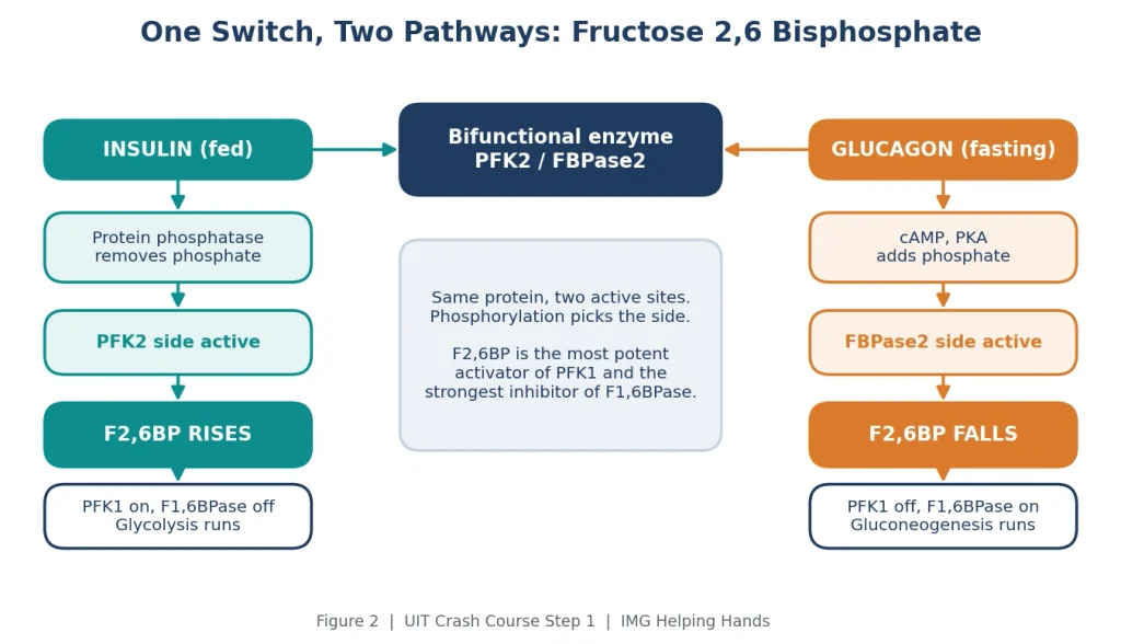
Figure 2. Insulin and glucagon act on the same bifunctional enzyme and push fructose 2,6 bisphosphate in opposite directions.
In the fed state insulin dephosphorylates the enzyme, the PFK2 side runs, F2,6BP rises, glycolysis goes and gluconeogenesis stops. In the fasting state glucagon raises cAMP, protein kinase A phosphorylates the enzyme, the FBPase2 side runs, F2,6BP falls, and the liver makes glucose. If a stem mentions a carbohydrate rich meal and asks about F2,6BP, the answer is always PFK1 on and F1,6BPase off.
Insulin signals the fed state and drives storage. Glucagon and epinephrine signal fasting or stress and drive breakdown. Glucagon works through cAMP and protein kinase A, which phosphorylates target enzymes. Insulin works through protein phosphatase 1, which removes the same phosphates. That means every enzyme in this table is the same enzyme in two states, and if you know the fed column you can derive the fasting column by flipping it.
Table 2 is the framework we ask students to reproduce from memory before they touch a question bank.
| Enzyme | Fed state (insulin, dephosphorylated) | Fasting state (glucagon, phosphorylated) |
|---|---|---|
| PFK1 | On, because F2,6BP is high | Off |
| Fructose 1,6 bisphosphatase | Off | On |
| Glycogen synthase | On | Off |
| Glycogen phosphorylase | Off | On |
| Acetyl CoA carboxylase | On | Off |
| CPT1 | Off, because malonyl CoA is high | On |
| Mitochondrial HMG CoA synthase | Off | On |
| HMG CoA reductase | On | Off |
| Hormone sensitive lipase (not rate limiting, same rule) | Off | On |
| UIT Exam Trap: Phosphorylation Does Not Mean Activation Students who learned phosphorylation as a generic on switch get half of these backwards. The rule is about direction, not activity. Phosphate turns on the enzymes that break things down (glycogen phosphorylase, hormone sensitive lipase) and turns off the enzymes that build things up (glycogen synthase, ACC, HMG CoA reductase). Glucagon adds the phosphate. Insulin takes it away. Say it that way and the table writes itself. |
Location is a quiet favorite of question writers because it separates the two carbamoyl phosphate synthetases and the two HMG CoA synthases. A rough rule that works when memory fails: catabolism that uses oxygen sits in the mitochondrion, synthesis that uses NADPH sits in the cytosol.
Mitochondrial: CPS I, isocitrate dehydrogenase, HMG CoA synthase for ketones, ALA synthase, and CPT1 on the outer mitochondrial membrane.
Cytosolic: PFK1, fructose 1,6 bisphosphatase, both glycogen enzymes, G6PD, CPS II, glutamine PRPP amidotransferase, acetyl CoA carboxylase, HMG CoA reductase and 7 alpha hydroxylase.
The exam almost never asks a bare enzyme question. It hides the enzyme inside a drug mechanism, a poisoning, or an inherited disease. These are the pairings that appear.
Table 3 lists the pharmacology and toxicology links, including the two that are traps because the agent does not hit the rate limiting enzyme.
| Drug or toxin | Enzyme affected | What the stem shows |
|---|---|---|
| Statins | HMG CoA reductase inhibited | LDL falls because hepatic LDL receptors increase. Myalgia and raised CK as the adverse effect |
| Cholestyramine, colestipol, colesevelam | 7 alpha hydroxylase increased (indirectly) | Bile acids lost in stool, liver converts more cholesterol to bile acids, LDL falls |
| 6 mercaptopurine, azathioprine | Glutamine PRPP amidotransferase inhibited | Immunosuppression, leukemia therapy. Allopurinol raises 6 MP levels |
| Lead | ALA dehydratase and ferrochelatase inhibited | Microcytic anemia, basophilic stippling, raised urine ALA. Not ALA synthase |
| Hemin and glucose | ALA synthase suppressed | Treatment of an acute intermittent porphyria attack |
| Barbiturates, other CYP inducers | ALA synthase induced | Precipitate porphyria attacks by consuming hepatic heme |
| Valproate | CPS I inhibited, NAG depleted | Hyperammonemia, sometimes with normal liver enzymes |
| Carglumic acid | CPS I activated (NAG analogue) | NAG synthase deficiency, some organic acidemias |
| Metformin (cross topic) | Gluconeogenesis suppressed via AMPK | Not a direct rate limiting enzyme inhibitor. Included so it is not confused with one |
Table 4 lists the diseases. Two of them, Von Gierke and OTC deficiency, involve enzymes downstream of the rate limiting step, but the exam files them in the same drawer.
| Disease | Enzyme | Hallmark |
|---|---|---|
| G6PD deficiency | Glucose 6 phosphate dehydrogenase | Hemolysis after oxidant drugs, fava beans or infection. Bite cells and Heinz bodies |
| McArdle disease (GSD V) | Muscle glycogen phosphorylase | Exercise cramps, no rise in lactate on forearm testing, second wind, myoglobinuria |
| Hers disease (GSD VI) | Liver glycogen phosphorylase | Mild fasting hypoglycemia and hepatomegaly |
| Von Gierke disease (GSD I) | Glucose 6 phosphatase (downstream) | Severe fasting hypoglycemia, lactic acidosis, hepatomegaly, hyperuricemia |
| Acute intermittent porphyria | Porphobilinogen deaminase, with ALA synthase induced upstream | Abdominal pain, neuropathy, psychiatric symptoms, urine darkens on standing |
| Lead poisoning | ALA dehydratase and ferrochelatase | Microcytic anemia, stippling, lead lines, neuropathy, raised urine ALA |
| Primary carnitine deficiency | Carnitine transport into cells, upstream of CPT1 | Weakness, cardiomyopathy, hypoketotic hypoglycemia |
| OTC deficiency | Ornithine transcarbamylase (downstream of CPS I) | Hyperammonemia with orotic aciduria, X linked |
Book a free call with mentors who matched as IMGs. We’ll build a Step 1 plan that trains this exact reflex across every subject, not just biochem.
We keep three. More than that and students spend the exam decoding mnemonics instead of answering questions.
CPS I is Inside, CPS II is Outside: CPS I sits inside the mitochondrion, runs the urea cycle, takes free ammonia and needs NAG. CPS II sits outside in the cytosol, runs pyrimidine synthesis, takes nitrogen from glutamine and is shut off by UTP. The Roman numeral tells you the compartment.
Phosphate Powers the Breakdown: phosphorylation activates breakdown enzymes and inactivates synthesis enzymes. Glucagon phosphorylates, insulin dephosphorylates. Every row in Table 2 follows from this one sentence.
Malonyl CoA is the traffic light: when ACC is making malonyl CoA the cell is building fat, and malonyl CoA blocks CPT1 so fat cannot enter the mitochondrion to be burned. Synthesis on means oxidation off. One molecule enforces it.
For the full list order, some students use a sentence built from the first letter of each pathway in First Aid order. It works if you already own the content, but it does not replace the fed versus fasting framework, and we have never seen a question that required reciting the list in order.
Nearly every stem in this category is one of eight scenarios. Figure 3 is the decision tree we use in class. Read the state or the exposure, then read the enzyme.
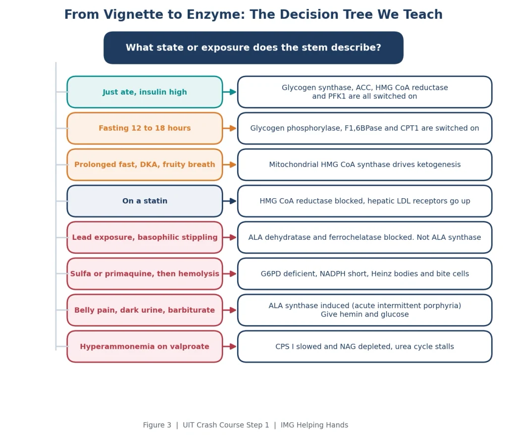
Figure 3. The eight scenarios that account for almost every rate limiting enzyme question, and the enzyme each one points to.
Ranked roughly by how often they decide the answer.
Six vignettes in NBME style. The wrong answer analysis matters more than the right answer, because the distractors are the enzymes you will confuse under pressure.
Difficulty: Easy | Competency: Biochemistry, fed and fasting regulation
A 22 year old medical student has not eaten for 14 hours while studying. Toward the end of this period her hepatic glucose output is maintained mainly by gluconeogenesis. Activity of which of the following enzymes is most likely increased compared with the fed state?
A. Phosphofructokinase 1
B. Acetyl CoA carboxylase
C. Fructose 1,6 bisphosphatase
D. HMG CoA reductase
E. Glycogen synthase
Correct answer: C. Fructose 1,6 bisphosphatase
Fourteen hours of fasting means glucagon is high. Glucagon raises cAMP, protein kinase A phosphorylates the bifunctional enzyme into its FBPase2 form, and fructose 2,6 bisphosphate falls. Without F2,6BP, PFK1 is inhibited and fructose 1,6 bisphosphatase is released from inhibition, driving gluconeogenesis.
Why the other options fail:
Difficulty: Easy | Competency: Pharmacology, cholesterol synthesis
A 45 year old man with hyperlipidemia is started on atorvastatin. Six weeks later his LDL cholesterol has fallen by 45 percent. Which of the following changes in his hepatocytes best explains the fall in serum LDL?
A. Increased activity of cholesterol 7 alpha hydroxylase
B. Increased expression of LDL receptors on the cell surface
C. Decreased activity of lipoprotein lipase
D. Increased activity of acetyl CoA carboxylase
E. Decreased secretion of VLDL
Correct answer: B. Increased expression of LDL receptors on the cell surface
Statins competitively inhibit HMG CoA reductase, the rate limiting enzyme of cholesterol synthesis. Hepatic cholesterol falls, the cell senses sterol depletion through SREBP, and LDL receptors are upregulated so the liver pulls more LDL out of the blood. The receptor change, not the enzyme block itself, is what lowers serum LDL.
Why the other options fail:
Difficulty: Medium | Competency: Biochemistry, heme synthesis and toxicology
A 4 year old boy has intermittent abdominal pain, irritability and difficulty concentrating. He lives in an apartment built in the 1950s with peeling paint. Hemoglobin is 9.2 g/dL with a low MCV, and the peripheral smear shows basophilic stippling. Urinary delta aminolevulinic acid is elevated. Which of the following enzymes is most likely inhibited?
A. ALA synthase
B. ALA dehydratase
C. Porphobilinogen deaminase
D. Uroporphyrinogen decarboxylase
E. HMG CoA reductase
Correct answer: B. ALA dehydratase
Lead inhibits ALA dehydratase and ferrochelatase. Blocking ALA dehydratase causes ALA to accumulate and spill into urine. Blocking ferrochelatase leaves protoporphyrin without iron, producing microcytic anemia and the ribosomal aggregates seen as basophilic stippling.
Why the other options fail:
Difficulty: Medium | Competency: Biochemistry, ketogenesis
A 35 year old man with type 1 diabetes presents with nausea, vomiting and fruity breath after running out of insulin three days ago. Plasma glucose is 480 mg/dL, arterial pH is 7.18 and the anion gap is 22. Serum beta hydroxybutyrate is markedly elevated. Activity of which of the following enzymes is most likely increased in his hepatocytes?
A. Acetyl CoA carboxylase
B. HMG CoA reductase
C. Mitochondrial HMG CoA synthase
D. Glycogen synthase
E. Pyruvate dehydrogenase
Correct answer: C. Mitochondrial HMG CoA synthase
Absent insulin and high glucagon release lipolysis in adipose tissue. Fatty acids flood the liver, beta oxidation generates acetyl CoA faster than the TCA cycle can use it, and mitochondrial HMG CoA synthase, the rate limiting enzyme of ketogenesis, converts the excess to ketone bodies.
Why the other options fail:
Difficulty: Hard | Competency: Biochemistry, reciprocal regulation of fat metabolism
A researcher generates a hepatocyte line in which the gene for acetyl CoA carboxylase has been deleted. The cells are grown in medium rich in glucose and amino acids. Compared with wild type cells, the mutant cells are most likely to show which of the following?
A. Increased malonyl CoA and decreased fatty acid oxidation
B. Decreased malonyl CoA and increased fatty acid oxidation
C. Increased malonyl CoA and increased fatty acid synthesis
D. Decreased malonyl CoA and decreased fatty acid oxidation
E. No change in either pathway
Correct answer: B. Decreased malonyl CoA and increased fatty acid oxidation
Acetyl CoA carboxylase makes malonyl CoA. Delete the enzyme and malonyl CoA disappears. Malonyl CoA is the inhibitor of CPT1, so without it CPT1 runs freely and long chain fatty acids enter the mitochondrion to be oxidized, even though the medium is signalling a fed state. This is the whole point of the malonyl CoA brake.
Why the other options fail:
Difficulty: Medium | Competency: Biochemistry and pharmacology, urea cycle
A 9 year old girl with epilepsy is started on valproic acid. Two weeks later she becomes lethargic and confused. Serum ammonia is markedly elevated while liver transaminases are normal. Which of the following best explains her hyperammonemia?
A. Inhibition of ornithine transcarbamylase
B. Depletion of N acetylglutamate with reduced carbamoyl phosphate synthetase I activity
C. Inhibition of carbamoyl phosphate synthetase II
D. Increased activity of glutaminase in the kidney
E. Hepatocellular necrosis with loss of urea cycle enzymes
Correct answer: B. Depletion of N acetylglutamate with reduced carbamoyl phosphate synthetase I activity
Valproate and its metabolites inhibit carbamoyl phosphate synthetase I directly and also deplete N acetylglutamate, the obligatory activator of CPS I. Without NAG the first step of the urea cycle stalls, ammonia is not captured, and encephalopathy follows. Normal transaminases tell you this is a metabolic effect, not liver injury.
Why the other options fail:
USMLE Impact Theory covers every high-yield topic with the same structure, NBME-style vignettes, wrong-answer analysis, and the traps written into the stems.
Yes. Pass or fail changed the consequences of a score, not the content of the exam. Biochemistry sits inside the Biochemistry and Nutrition section and the Multisystem Processes and Disorders section of the content outline, and metabolic regulation is a large part of it. A borderline candidate who loses an entire biochemistry block can fail. Rate limiting enzymes are the most efficient part of that block to learn because a small list covers a lot of questions.
Yes, but the list is smaller than it looks. The regulators repeat: insulin and glucagon, ATP and AMP, citrate, and end product feedback account for almost every entry. Learn the fed versus fasting framework once and the master table follows. The only enzymes that need separate memorization are the nitrogen and heme enzymes, because they answer to feedback rather than hormones.
CPS I is in the mitochondrion, uses free ammonia, needs N acetylglutamate and starts the urea cycle. CPS II is in the cytosol, uses glutamine, is activated by ATP and PRPP, is inhibited by UTP and starts pyrimidine synthesis. They are different genes and different proteins that happen to share a name. Mixing them up is the single most common error in this category.
Because one molecule controls two opposing pathways. It activates PFK1 and inhibits fructose 1,6 bisphosphatase, so its level alone tells you whether the liver is burning glucose or making it. Question writers like it because a single stem about F2,6BP tests glycolysis, gluconeogenesis, insulin signalling and glucagon signalling at once.
Students learn that ALA synthase is the rate limiting enzyme of heme synthesis, then see a heme synthesis question and pick it. Lead does not act there. It inhibits ALA dehydratase and ferrochelatase. The clue is elevated urinary ALA: if ALA synthase were blocked, ALA would be low, not high.
Blocking HMG CoA reductase lowers cholesterol inside the hepatocyte. The cell responds to that shortage by putting more LDL receptors on its surface and pulling LDL out of the blood. The receptor upregulation is the mechanism that moves serum LDL, and it is the step the exam usually asks about.
Malonyl CoA is the product of acetyl CoA carboxylase, the first committed step of fatty acid synthesis. It also inhibits CPT1, the enzyme that lets fatty acids into the mitochondrion for oxidation. So whenever the cell is building fat it is also blocking fat burning. Questions about why a fed liver does not oxidize fatty acids, or what happens if ACC is deleted, are all this one connection.
Muscle has no glucagon receptors. Its glycogen phosphorylase responds to AMP, calcium and epinephrine, and the glucose it releases stays in the muscle because muscle lacks glucose 6 phosphatase. That is why McArdle disease presents with exercise intolerance rather than fasting hypoglycemia, and why liver glycogen phosphorylase deficiency, Hers disease, looks completely different.
Both suppress ALA synthase. Hemin restores the heme pool that feedback inhibits the enzyme. Glucose represses its transcription, which is also why fasting precipitates attacks. Drugs that induce cytochrome P450 do the opposite: they consume heme, lift the feedback, and set off an attack.
Draw Figure 1 from memory, then Table 2. If you can do both without looking, you have the framework. After that, run the eight scenarios in Figure 3 against a question bank and keep a list of every enzyme you confuse. Most students find the list has three or four entries and they are always the same ones.
This is exactly how USMLE Impact Theory works, built by IMGs, for IMGs, on the same one-framework-then-every-vignette engine you just used to line up fifteen enzymes. Here’s what that looks like across the whole exam:
Articles published by IMG Helping Hands are prepared by our team using information from direct experience, publicly available resources, and educational references. AI tools may be used to assist with drafting, proofreading, and formatting; however, all content undergoes review and approval before publication.
The information provided is intended for educational purposes only. Requirements, policies, and processes may change over time. Readers should consult official sources for the most current information.