
| TORCH infections are a group of congenital infections that cross from mother to fetus and cause overlapping newborn findings such as growth restriction, rash, hearing loss, and intracranial calcifications. The acronym stands for Toxoplasma, Other (syphilis, Listeria, parvovirus B19, VZV, HIV, and Zika), Rubella, Cytomegalovirus, and Herpes simplex virus. Each bug is separated on exams by one discriminating feature, for example diffuse calcifications in toxoplasmosis versus periventricular calcifications in CMV. |
Congenital bugs feel confusing because their pictures overlap. The trick that makes TORCH infections easier is to anchor each organism to a single discriminator and a single buzzword. That is exactly how this guide is built, and it is the pattern recognition method IMG Helping Hands uses inside the USMLE Impact Theory (UIT) course to train International Medical Graduates.
TORCH is an acronym for the classic congenital infections that share a common route (vertical transmission from mother to fetus) and a common set of newborn findings. What does TORCH stand for, letter by letter:
| Letter | Stands for | Notes |
|---|---|---|
| T | Toxoplasma gondii | Diffuse intracranial calcifications, chorioretinitis, hydrocephalus |
| O | Other | Syphilis, Listeria, Parvovirus B19, VZV, HIV, and Zika. This is where exams trip people. |
| R | Rubella | Congenital rubella syndrome, the three Cs (cataracts, cardiac, cochlear) |
| C | Cytomegalovirus | Most common congenital infection, periventricular calcifications, hearing loss |
| H | Herpes simplex virus (and HIV) | Perinatal transmission, vesicles, encephalitis |
The letter that decides exam questions is O for Other, because it hides six high yield bugs. Modern Step 1 items expect you to include Zika here alongside the traditional agents.
| MNEMONIC: The Other bugs, Silly Little Parasites Vex Happy Zygotes: Syphilis, Listeria, Parvovirus B19, VZV, HIV, Zika. Keep this string ready, because a maternal exposure clue (a cat, mosquito travel, deli meat, a facial rash) is often the only hint the vignette gives. |
| TRAP: Do not read the O as a single disease. It is a bucket of six organisms, and syphilis alone accounts for a large share of congenital infection vignettes. If a stem lists snuffles, a saddle nose, or notched teeth, the answer is in the Other group, not Toxoplasma or Rubella. |
Before you reach for a discriminator, recognize the common picture that almost all of these infections share. Any TORCH infection can produce intrauterine growth restriction, hepatosplenomegaly, thrombocytopenia with petechiae, jaundice, and a blueberry muffin rash. These overlapping findings are why the bugs feel confusing. The strategy is to spot the shared picture first, then let one discriminating feature name the exact organism.
| HIGH YIELD: When several of these appear together in a newborn (growth restriction, hepatosplenomegaly, petechiae, jaundice, blueberry muffin rash), think congenital infection first, then use the discriminator (calcification pattern, eye finding, or maternal exposure) to pick the organism. |
Vertical transmission means spread from mother to child. There are three routes. Transplacental spread happens in utero, when the organism crosses the placenta. Perinatal spread happens at delivery, when the newborn contacts infected genital secretions. Breast milk spread happens after birth, for example with CMV and HIV.
Timing changes the outcome. As a rule, earlier infection produces more severe congenital disease because it disrupts organ formation, while later infection carries a higher chance of transmission. Rubella is the classic first trimester teratogen. HSV is the outlier, because it is usually caught at delivery rather than in the womb.
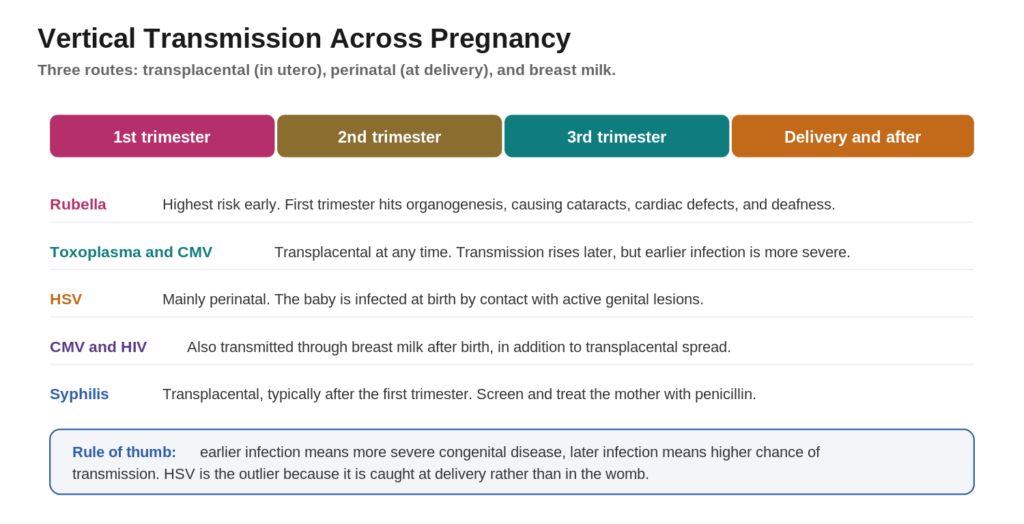
| HIGH YIELD: Two questions answer most transmission items. When was the mother infected, and by what route. First trimester rubella gives the worst structural defects. HSV and perinatal exposure to active lesions drive the delivery decision, which is cesarean section when active genital lesions are present. |
A TORCH panel measures antibodies against the classic agents. The concept that turns this into a test question is antibody biology. Maternal IgG crosses the placenta, but IgM does not. So a positive IgG in the newborn may simply be borrowed from the mother, while a raised IgM in the newborn was made by the baby and points to a true congenital infection. Confirmation then depends on the organism, for example PCR for CMV, dark field microscopy and serology for syphilis, and organism specific serology for Toxoplasma and Rubella.
| HIGH YIELD: Newborn IgM means the infant made it, because IgM cannot cross the placenta. A raised neonatal IgM is the classic clue that a congenital infection is real rather than transferred maternal IgG. |
This is the centerpiece. One row per bug, built for fast recall and for direct citation. Use it as your TORCH infections chart.
| Infection | Organism | Transmission and timing | Classic triad and buzzwords | Distinguishing finding and maternal clue | Prevention and treatment |
|---|---|---|---|---|---|
| Toxoplasmosis | Toxoplasma gondii | Transplacental, any trimester | Chorioretinitis, hydrocephalus, diffuse calcifications | DIFFUSE calcifications. Cat litter or undercooked meat | Avoid cat litter and raw meat. Spiramycin for mother, pyrimethamine plus sulfadiazine plus leucovorin for infant |
| Rubella | Rubella virus | Transplacental, worst in first trimester | Cataracts, cardiac defects, deafness, blueberry muffin rash | PDA and pulmonary artery stenosis, cataracts. Unvaccinated mother with a rash | MMR vaccine before pregnancy. Supportive care |
| CMV | Cytomegalovirus | Transplacental, perinatal, and breast milk | Periventricular calcifications, hearing loss, petechiae, owl eye inclusions | PERIVENTRICULAR calcifications, sensorineural hearing loss. Often asymptomatic mother | Ganciclovir or valganciclovir for symptomatic infants |
| Herpes (HSV) | HSV 2 mainly | Perinatal, at delivery | Skin, eye, and mouth vesicles, encephalitis | Temporal lobe encephalitis, vesicles. Active genital lesions | Cesarean section if active lesions. Acyclovir |
| Syphilis | Treponema pallidum | Transplacental, usually after first trimester | Snuffles, saddle nose, Hutchinson teeth, saber shins | Notched Hutchinson teeth, mulberry molars, saber shins. Positive maternal RPR or VDRL | Screen and treat mother with penicillin |
| Listeriosis | Listeria monocytogenes | Transplacental and perinatal | Granulomatosis infantiseptica, sepsis | Widespread microabscesses and granulomas. Unpasteurized dairy or deli meat | Ampicillin |
| Parvovirus B19 | Parvovirus B19 | Transplacental | Hydrops fetalis, severe anemia | Nonimmune hydrops from fetal aplastic anemia. Mother with a lacy facial rash | Intrauterine transfusion if severe |
| Varicella (VZV) | Varicella zoster virus | Transplacental | Limb hypoplasia, cicatricial skin scarring | Dermatomal scars and limb defects. Maternal chickenpox in early pregnancy | VZIG and vaccination before pregnancy |
| HIV | Human immunodeficiency virus | Transplacental, perinatal, breast milk | Often asymptomatic at birth, later infections | Failure to thrive and recurrent infections | Maternal antiretroviral therapy greatly reduces transmission |
| Zika | Zika virus | Transplacental, Aedes mosquito | Microcephaly, intracranial calcifications | Severe microcephaly with brain disruption. Mosquito exposure or travel | Avoid travel to endemic areas, mosquito precautions |
That’s the UIT method. Learn every high-yield Step 1 microbiology topic as pattern recognition, not memorization.
Toxoplasma gondii crosses the placenta after a primary maternal infection. The classic triad is chorioretinitis, hydrocephalus, and diffuse intracranial calcifications. The maternal clue is exposure to cat feces during litter box changes or eating undercooked meat. The parasite spreads as tachyzoites and persists in tissue as cysts of bradyzoites.
| HIGH YIELD: Toxoplasmosis calcifications are DIFFUSE and scattered throughout the brain. Pair that with chorioretinitis and hydrocephalus and the diagnosis is locked. |
Rubella is the classic first trimester teratogen. Congenital rubella syndrome is remembered as the three Cs: Cataracts, Cardiac defects (patent ductus arteriosus and pulmonary artery stenosis), and Cochlear defects (sensorineural deafness). A salt and pepper retinopathy and a blueberry muffin rash may also be present. Rubella is a togavirus, and the maternal clue is an unvaccinated mother with a viral rash in early pregnancy.
| MNEMONIC: Congenital rubella, the three Cs: Cataracts, Cardiac (PDA), Cochlear (sensorineural deafness). If a newborn has cataracts plus a continuous murmur plus deafness, think rubella first. |
Cytomegalovirus is the most common congenital infection and the leading nongenetic cause of sensorineural hearing loss. Findings include periventricular calcifications, sensorineural hearing loss, petechiae, hepatosplenomegaly, and microcephaly. CMV is a herpesvirus, and infected cells show characteristic owl eye intranuclear inclusions on histology. Many mothers are asymptomatic, which is why CMV is easy to miss.
| TRAP: The single most tested contrast in this whole topic is Toxo versus CMV calcifications. CMV calcifications hug the ventricles (periventricular). Toxoplasma calcifications are spread diffusely. Reversing these two is the most common careless error on Step 1. |
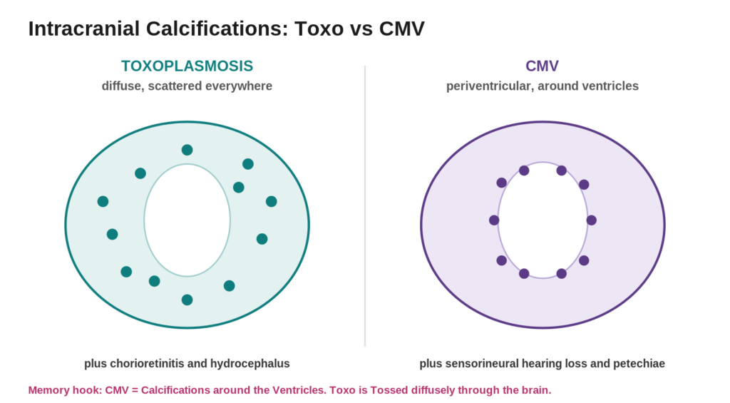
| MNEMONIC CMV = Calcifications around the Ventricles. Both start with a V sound. Toxo is Tossed diffusely through the brain. |
Neonatal herpes, usually HSV 2, is acquired perinatally during passage through an infected birth canal, especially with active lesions. Disease appears in the first weeks of life as skin, eye, and mouth vesicles, as central nervous system disease with temporal lobe encephalitis, or as disseminated infection. Delivery by cesarean section is recommended when active genital lesions are present, and treatment is acyclovir. Risk is highest when the mother has a primary HSV infection near term, and fetal scalp electrodes should be avoided during labor because they can inoculate the virus.
| HIGH YIELD: Unlike the transplacental TORCH bugs, HSV is a delivery event. A newborn with vesicles and seizures, born vaginally to a mother with genital lesions, is neonatal herpes until proven otherwise. |
Treponema pallidum crosses the placenta. Early signs include snuffles (an infectious nasal discharge), a maculopapular rash on the palms and soles, and hepatosplenomegaly. Late stigmata include notched Hutchinson teeth, mulberry molars, a saddle nose, saber shins, and sensorineural deafness. Maternal screening uses nontreponemal tests (RPR or VDRL), confirmed by treponemal tests (FTA ABS or TP antibody), and the spirochete can be seen on dark field microscopy.
| MNEMONIC: Congenital syphilis stigmata: the S cluster of Snuffles, Saddle nose, and Saber shins, plus the Hutchinson triad of Hutchinson teeth, interstitial keratitis, and eighth nerve deafness. Screen and treat the mother with penicillin to prevent all of it. |
Listeria monocytogenes causes granulomatosis infantiseptica, a disseminated infection with widespread microabcesses, linked to unpasteurized dairy and deli meats. Parvovirus B19 causes hydrops fetalis through fetal aplastic anemia, with the maternal clue of a lacy facial rash. VZV in early pregnancy causes limb hypoplasia and cicatricial dermatomal scarring. congenital HIV and perinatal transmission and is prevented by maternal antiretroviral therapy. Zika, spread by Aedes mosquitoes, causes severe microcephaly with intracranial calcifications.
A blueberry muffin rash is made of blue purple skin nodules that represent dermal extramedullary hematopoiesis, that is, blood cell production in the skin. Infectious causes are mainly rubella and CMV, along with toxoplasmosis, parvovirus B19, and syphilis. Noninfectious causes include congenital leukemia, neuroblastoma, hemolytic disease of the newborn (Rh), twin to twin transfusion, and hereditary spherocytosis.
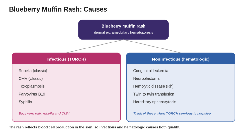
| MNEMONIC: Blueberry muffin, two buzzword bugs plus a blood bucket: infectious equals Rubella and CMV first, then Toxo, Parvovirus, and Syphilis. Noninfectious equals hematologic, such as leukemia, neuroblastoma, and hemolytic disease. The rash is blood forming in the skin, so both categories fit. |
Two patterns matter. Toxoplasma produces diffuse calcifications scattered across the brain. CMV produces periventricular calcifications lining the ventricles. Zika tends to produce subcortical or junctional calcifications with striking microcephaly.
| Pattern | Infection | Best partner clue |
|---|---|---|
| Diffuse, scattered | Toxoplasmosis | Chorioretinitis and hydrocephalus, cat or raw meat exposure |
| Periventricular | CMV | Sensorineural hearing loss and petechiae, often asymptomatic mother |
| Subcortical with microcephaly | Zika | Mosquito exposure or travel to an endemic area |
An eye finding narrows the field fast. Cataracts (a white cloudy lens, often with a cardiac murmur and deafness) point to rubella. Chorioretinitis (inflammation of the retina and choroid) points to toxoplasmosis or CMV. Cataracts are structural and early, chorioretinitis is inflammatory.
Both CMV and rubella cause sensorineural hearing loss. CMV is the leading nongenetic cause of congenital sensorineural hearing loss overall, and the loss can appear or progress after birth. Rubella hearing loss travels with cataracts and cardiac defects. Use the companion findings to break the tie.
| Buzzword or clue | Diagnosis |
|---|---|
| Diffuse intracranial calcifications, cat litter | Congenital toxoplasmosis |
| Periventricular calcifications, petechiae | Congenital CMV |
| Owl eye intranuclear inclusions | Congenital CMV |
| Number one nongenetic cause of hearing loss | Congenital CMV |
| Most common congenital infection | Congenital CMV |
| Cataracts plus PDA plus deafness (three Cs) | Congenital rubella syndrome |
| Salt and pepper retinopathy | Congenital rubella syndrome |
| Raised neonatal IgM (does not cross placenta) | Confirms a true congenital infection |
| Blueberry muffin rash, two classic bugs | Rubella and CMV |
| Vesicles and temporal lobe encephalitis in a newborn | Neonatal HSV |
| Snuffles, saddle nose, Hutchinson teeth, saber shins | Congenital syphilis |
| Granulomatosis infantiseptica, deli meat | Congenital Listeria |
| Hydrops fetalis, lacy facial rash in mother | Parvovirus B19 |
| Limb hypoplasia, dermatomal skin scars | Congenital varicella (VZV) |
| Severe microcephaly, mosquito exposure | Congenital Zika |
TORCH infections are a group of congenital infections that pass from mother to fetus and cause overlapping newborn problems such as growth restriction, rash, hearing loss, and intracranial calcifications. The letters stand for Toxoplasma, Other (syphilis, Listeria, parvovirus B19, VZV, HIV, Zika), Rubella, Cytomegalovirus, and Herpes simplex virus.
The O stands for Other, a bucket of six important organisms: syphilis, Listeria, parvovirus B19, varicella zoster virus, HIV, and Zika. Modern exams expect Zika to be included.
Look at the calcification pattern. Toxoplasmosis causes diffuse scattered calcifications and pairs with chorioretinitis and hydrocephalus. CMV causes periventricular calcifications and pairs with sensorineural hearing loss and petechiae.
The rash reflects blood cell production in the skin. The classic infectious causes are rubella and CMV, along with toxoplasmosis, parvovirus B19, and syphilis. Noninfectious causes include congenital leukemia, neuroblastoma, and hemolytic disease of the newborn.
A TORCH panel screens for antibodies against the classic congenital agents. The key rule is that maternal IgG crosses the placenta but IgM does not, so a raised IgM in the newborn indicates a true congenital infection rather than transferred maternal antibody. Confirmation depends on the organism, such as PCR for CMV, dark field microscopy and serology for syphilis, and specific serology for Toxoplasma and Rubella.
CMV is the most common congenital infection and the leading nongenetic cause of sensorineural hearing loss. It matters because many mothers are asymptomatic, so affected newborns can be missed without screening for hearing loss.
See the highest-yield Step 1 systems mapped the same discriminator-first way, free, and built for IMGs.
Original questions written in Step 1 clinical vignette format. Answers and explanations follow.
A newborn has a head circumference below the third percentile. Head imaging shows periventricular calcifications, and the infant fails the newborn hearing screen. The mother recalls only a mild flu like illness in pregnancy. Which organism is most likely responsible?
A. Toxoplasma gondii
B. Cytomegalovirus
C. Rubella virus
D. Treponema pallidum
E. Herpes simplex virus
A pregnant woman who gardens and keeps several cats delivers an infant with hydrocephalus, chorioretinitis, and diffuse intracranial calcifications scattered throughout the brain. Which exposure most likely led to this infection?
A. Contact with cat feces or undercooked meat
B. Passage through an infected birth canal
C. A mosquito bite while traveling
D. Eating unpasteurized soft cheese
E. Receiving a blood transfusion
A newborn has bilateral cataracts, a continuous machine like murmur, and fails the hearing screen. The mother immigrated recently and never received childhood vaccinations. Which finding best explains the murmur?
A. Ventricular septal defect
B. Patent ductus arteriosus
C. Coarctation of the aorta
D. Tetralogy of Fallot
E. Atrial septal defect
A 6 day old infant born by vaginal delivery develops clusters of vesicles on the scalp, then seizures. MRI shows temporal lobe involvement. The mother had painful genital lesions near her due date. What is the most appropriate treatment?
A. Penicillin G
B. Acyclovir
C. Ganciclovir
D. Pyrimethamine and sulfadiazine
E. Ampicillin
A newborn has a persistent nasal discharge (snuffles), a maculopapular rash on the palms and soles, and hepatosplenomegaly. Radiographs later show saber shins. Which maternal test would most likely have been positive during pregnancy?
A. Rubella IgG titer
B. RPR or VDRL
C. CMV PCR
D. Toxoplasma serology
E. HIV viral load
A term newborn appears well but the pediatrician wants to prevent the most common cause of nongenetic sensorineural hearing loss from being missed. Which congenital infection is the target of this concern?
A. Rubella
B. CMV
C. Toxoplasmosis
D. Syphilis
E. Parvovirus B19
A fetus is found on ultrasound to have scalp edema, pleural effusions, and ascites (hydrops fetalis) with severe anemia. Two weeks earlier the mother, a schoolteacher, had a lacy reticular rash on her arms and a slapped cheek appearance. Which agent is responsible?
A. Parvovirus B19
B. Cytomegalovirus
C. Listeria monocytogenes
D. Rubella virus
E. Zika virus
A newborn has a blueberry muffin rash, and initial TORCH serologies are being processed. Which pair of infections is classically associated with this rash?
A. Rubella and CMV
B. HSV and HIV
C. Listeria and syphilis
D. Zika and VZV
E. Toxoplasma and parvovirus
A pregnant woman returns from travel to a region with active mosquito borne transmission. Her fetus is later found to have severe microcephaly with intracranial calcifications and a collapsed skull appearance. Which infection is most likely?
A. Zika virus
B. Rubella virus
C. Toxoplasma gondii
D. Cytomegalovirus
E. Syphilis
A woman with painful active genital herpes lesions is in early labor at term. Which intervention most reduces the risk of neonatal herpes?
A. Oral acyclovir for the newborn only
B. Cesarean section delivery
C. Immediate breastfeeding
D. Intramuscular penicillin for the mother
E. Maternal antiretroviral therapy
1. Correct answer B. Periventricular calcifications plus sensorineural hearing loss plus microcephaly, with an asymptomatic or mildly ill mother, is classic congenital CMV. Toxoplasma gives diffuse calcifications, and rubella pairs deafness with cataracts and cardiac defects.
2. Correct answer A. Cat feces and undercooked meat transmit Toxoplasma gondii, which causes the triad of chorioretinitis, hydrocephalus, and diffuse calcifications. Birth canal passage is HSV, mosquito travel is Zika, soft cheese is Listeria.
3. Correct answer B. Congenital rubella syndrome features the three Cs, and the cardiac lesion is a patent ductus arteriosus (a continuous machine like murmur), often with pulmonary artery stenosis. The other defects do not fit the cataract and deafness picture.
4. Correct answer B. Neonatal HSV with vesicles and temporal lobe encephalitis is treated with acyclovir. Penicillin treats syphilis, ganciclovir treats CMV, pyrimethamine and sulfadiazine treat toxoplasmosis, and ampicillin treats Listeria.
5. Correct answer B. Snuffles, a palm and sole rash, hepatosplenomegaly, and saber shins are congenital syphilis, so the maternal RPR or VDRL would be positive. The other tests do not explain saber shins and snuffles.
6. Correct answer B. CMV is both the most common congenital infection and the leading nongenetic cause of sensorineural hearing loss, and infected newborns are often asymptomatic, so hearing must be screened and followed.
7. Correct answer A. Parvovirus B19 infects erythroid precursors and causes fetal aplastic anemia leading to hydrops fetalis. The maternal slapped cheek and lacy rash of erythema infectiosum is the giveaway. CMV and rubella do not classically cause pure aplastic hydrops.
8. Correct answer A. Rubella and CMV are the two classic infectious causes of a blueberry muffin rash, which reflects dermal extramedullary hematopoiesis. The other pairs are not the classic association.
9. Correct answer A. Severe microcephaly with intracranial calcifications after mosquito borne exposure is congenital Zika. Rubella and CMV cause less severe microcephaly, and their calcification patterns and companion findings differ.
10. Correct answer B. Cesarean section delivery avoids contact with active genital lesions and most reduces neonatal HSV transmission. Penicillin is for syphilis and antiretroviral therapy is for HIV, neither of which addresses HSV at delivery.
Get an honest read on your Step 1 prep from IMG mentors who’ve matched, and a plan built around your weak areas.
TORCH infections stop being confusing the moment you attach one discriminator to each bug. Diffuse calcifications mean Toxoplasma. Periventricular calcifications mean CMV. The three Cs mean Rubella. Vesicles and temporal lobe encephalitis mean HSV. Snuffles and saber shins mean syphilis. Layer the maternal exposure clue on top and the newborn vignette solves itself.
IMG HELPING HANDS – UIT USMLE STEP 1 PROGRAM
Diffuse calcifications mean Toxoplasma. Periventricular means CMV. The three Cs mean rubella. One feature per organism, and a wall of overlapping vignettes turns into a matching exercise. That’s the entire USMLE Impact Theory method, concept first, discriminator second, buzzword last, anchored with visual memory.
The same logic runs through every high-yield system on Step 1. UIT crash courses teach microbiology, pharmacology, pathology, and physiology this way, live, mapped to First Aid, and built specifically for IMGs.
Pattern-based teaching. Mnemonic-driven recall. FA-mapped structure. Live IMG mentorship from doctors who’ve matched.
Learn the discriminator once. Recognize the bug every time.
Note: Educational content for exam preparation. It is not a substitute for clinical judgment or primary references.
Articles published by IMG Helping Hands are prepared by our team using information from direct experience, publicly available resources, and educational references. AI tools may be used to assist with drafting, proofreading, and formatting; however, all content undergoes review and approval before publication.
The information provided is intended for educational purposes only. Requirements, policies, and processes may change over time. Readers should consult official sources for the most current information.