
A hypersensitivity reaction is an exaggerated or inappropriate immune response to an antigen that results in tissue damage rather than protection. The immune system, which normally defends against pathogens, misfires and attacks the body’s own tissues, harmless environmental substances, or therapeutic agents.
The Gell and Coombs classification divides these reactions into four types based on the immune mechanism involved. Types I, II, and III are antibody mediated. Type IV is the only type mediated entirely by T cells, with no antibody involvement. This classification is the backbone of how USMLE tests immunopathology.
| Why This Matters in 2026 Even though USMLE Step 1 is now pass/fail, immunology remains critical. These concepts directly feed into Step 2 CK, clinical clerkships, and shelf exams.Hypersensitivity questions appear across immunology, pathology, pharmacology, and organ system blocks. Expect 4 to 6 questions per exam form.Strong immunology fundamentals also build the clinical reasoning you need for residency interviews and board certification. |
Here is the truth that nobody tells you. Hypersensitivity reactions are not hard because the science is complicated. They are hard because NBME writers deliberately blur the lines between types, hide the mechanism inside a clinical vignette, and give you four answer choices that all sound partially correct.
Most IMGs rely on a single First Aid table and assume memorizing “Type I equals IgE” is enough. It is not. The USMLE tests pattern recognition, not pattern recall. You need to identify a Type II reaction when the stem never mentions antibodies. You need to spot Type III when the question reads like rheumatology.
That is exactly how UIT at IMG Helping Hands teaches immunology. We do not ask you to memorize lists. We train you to read vignettes the way examiners write them. This guide breaks down every hypersensitivity type with the same clinical logic, flowcharts, mnemonics, and NBME traps we use in live mentorship sessions.
Before diving into each type, anchor this master table in your memory. Every USMLE hypersensitivity question can be solved by mapping the vignette back to one row of this table.
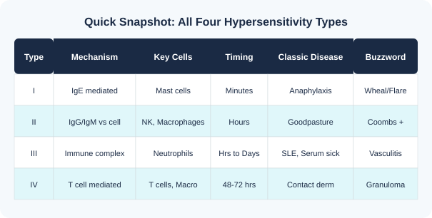
| Type | Alternate Name | Antibody | Key Mediators | Onset | Classic Example |
| I | Immediate / Anaphylactic | IgE | Histamine, Leukotrienes | Seconds to minutes | Anaphylaxis, Allergic asthma |
| II | Cytotoxic / Antibody mediated | IgG, IgM | Complement, NK cells | Hours | AIHA, Goodpasture, Graves |
| III | Immune complex | IgG (mainly) | Complement, Neutrophils | Hours to days | SLE, Serum sickness, Arthus |
| IV | Delayed type | None (T cells) | Cytokines, Macrophages | 48 to 72 hours | Contact dermatitis, TB test |
Hypersensitivity is not a single topic. It is a framework connecting immunology to every organ system. Examiners love it because one concept can be tested through pathology, pharmacology, rheumatology, nephrology, pulmonology, and dermatology.

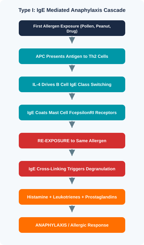
Type I hypersensitivity is an immediate, IgE mediated allergic reaction. It is the fastest of all four types. Mast cells and basophils are pre-sensitized with IgE antibodies, and re-exposure to the same allergen triggers rapid degranulation within seconds to minutes. Type I hypersensitivity reactions include anaphylaxis, allergic asthma, allergic rhinitis, and food allergies.
| Disease | Key Feature | NBME Buzzword |
| Anaphylaxis | Systemic IgE response, hypotension, laryngeal edema | Bee sting + throat swelling + hypotension |
| Allergic asthma | Bronchospasm, eosinophils in sputum, reversible obstruction | Wheezing after allergen exposure, responds to albuterol |
| Allergic rhinitis | Nasal congestion, sneezing, pale boggy turbinates | Seasonal symptoms, “allergic shiners” |
| Atopic dermatitis | Eczema, elevated IgE, flexural distribution in children | Itchy rash in antecubital fossae, family history of atopy |
| Food allergy | Urticaria, GI symptoms, can progress to anaphylaxis | Lip swelling minutes after peanut exposure |
| Drug allergy (acute) | Urticaria or anaphylaxis within minutes of drug | Rash minutes after penicillin injection |
| Eosinophilic esophagitis | Dysphagia, eosinophilic infiltration of esophageal mucosa | Young male with food impaction, rings on endoscopy |
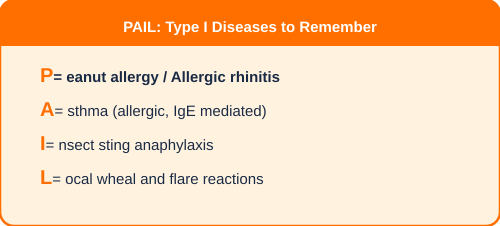
| NBME Trap Alert: Type I A stem describing anaphylaxis will NOT always say “IgE.” Look for: rapid onset, known allergen, urticaria, throat tightness, hypotension.Atopic triad (asthma + eczema + allergic rhinitis) is a classic vignette setup. If all three appear, the mechanism is Type I.Drug allergy timing matters: minutes = Type I. Days = think Type III (serum sickness).Serum tryptase confirms mast cell degranulation. When NBME includes this lab, the answer involves mast cells.Do not confuse allergic asthma (Type I, IgE) with hypersensitivity pneumonitis (Type III/IV, no IgE). |
Type I = IgE + Mast cells + Minutes + Histamine = Anaphylaxis spectrum.

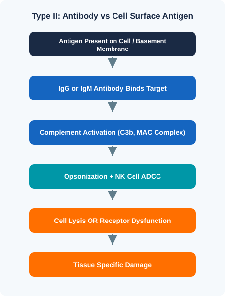
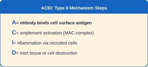
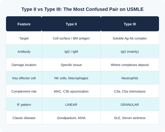
Type II hypersensitivity involves IgG or IgM antibodies directed against antigens on cell surfaces or basement membranes. Antibody binding triggers complement activation, opsonization, or antibody dependent cellular cytotoxicity (ADCC) by NK cells. The damage is tissue specific, which is the key difference from Type III where damage depends on where immune complexes deposit.
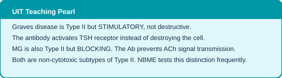
Type II is not always cytotoxic. USMLE specifically tests that some Type II reactions stimulate or block receptors without destroying the target cell.
| Subtype | Mechanism | Classic Example |
| Cytotoxic (Cell Destroying) | Complement MAC + ADCC destroys target cell | AIHA, Goodpasture, Transfusion reactions, HDN |
| Opsonization | C3b tags cells for phagocytosis by macrophages | Immune thrombocytopenia (ITP) |
| Functional (Non-Cytotoxic) | Antibody stimulates or blocks receptor function | Graves (stimulatory), Myasthenia gravis (blocking) |
| Disease | Target Antigen | Mechanism | Key Finding |
| Goodpasture syndrome | Type IV collagen (GBM + ABM) | Anti-GBM antibodies | Linear IF, hemoptysis + hematuria |
| Autoimmune hemolytic anemia | RBC surface antigens | IgG or IgM vs RBCs | Positive direct Coombs test, spherocytes |
| Graves disease | TSH receptor | Stimulatory IgG antibody | Hyperthyroidism, exophthalmos, diffuse goiter |
| Myasthenia gravis | AChR at NMJ | Blocking IgG antibody | Fatigable weakness, ptosis, improves with rest |
| Rheumatic fever | Cardiac myosin (molecular mimicry) | Anti-streptococcal Ab cross-react | Jones criteria, Aschoff bodies, pancarditis |
| Hemolytic disease of newborn | Rh antigen on fetal RBCs | Maternal IgG crosses placenta | Jaundice, hydrops fetalis, kernicterus |
| Pemphigus vulgaris | Desmoglein 3 (desmosomes) | Anti-desmosome IgG | Flaccid blisters, positive Nikolsky sign |
| Bullous pemphigoid | BP180/BP230 (hemidesmosomes) | Anti-hemidesmosome IgG | Tense blisters, negative Nikolsky sign |
| Acute transplant rejection (humoral) | Donor endothelium | Preformed donor-specific Ab | Rapid graft failure, C4d staining |
Type II = IgG/IgM against cell surface or basement membrane + Complement/ADCC + Tissue specific damage or receptor dysfunction.

Type III hypersensitivity occurs when soluble antigen-antibody immune complexes form in the circulation, deposit in tissues, activate complement, and recruit neutrophils that cause inflammation and tissue damage. The damage is NOT tissue specific. It depends entirely on WHERE the complexes deposit, most commonly in blood vessel walls, glomeruli, joints, and skin.
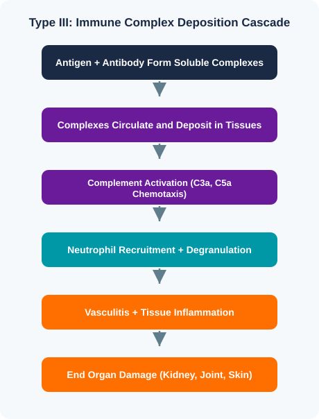
Immune complex deposition is not random. Complexes preferentially lodge in tissues with high blood flow and filtration: renal glomeruli (fenestrated capillaries), synovial joints, and dermal vessels. This explains the classic triad of nephritis, arthritis, and skin rash seen in SLE and serum sickness. Small immune complexes evade clearance and deposit more readily than large ones, which are efficiently removed by the reticuloendothelial system.
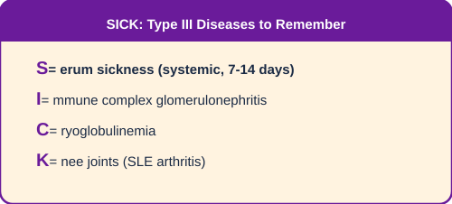
| Disease | Antigen Source | Deposition Site | Key Finding |
| SLE (lupus nephritis) | Anti-dsDNA, anti-Smith | Kidney, skin, joints, serosal surfaces | Granular IF, wire loop lesion (Class IV) |
| Post-streptococcal GN | Streptococcal antigens | Glomeruli (subepithelial) | Subepithelial humps on EM, “starry sky” IF |
| Serum sickness | Foreign proteins (drugs, antithymocyte globulin) | Systemic (vessels, kidney, joints) | Fever, urticaria, arthralgia, proteinuria 7-14d post drug |
| Arthus reaction | Locally injected antigen | Injection site only | Local edema, hemorrhage, necrosis within hours |
| Polyarteritis nodosa | Hepatitis B antigen | Medium vessel walls | Fibrinoid necrosis, string of beads on angiography |
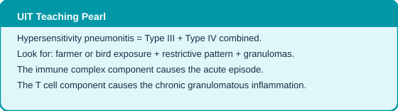
| Hypersensitivity pneumonitis | Inhaled organic dust (bird droppings, hay) | Lung interstitium | Farmer lung, bird fancier lung, restrictive PFTs |
| IgA vasculitis (HSP) | IgA immune complexes | Skin, joints, GI, kidney | Palpable purpura on buttocks/legs, child after URI |
| Mixed cryoglobulinemia | Hep C associated immune complexes | Skin, joints, kidney | Palpable purpura, low complement, Hep C positive |
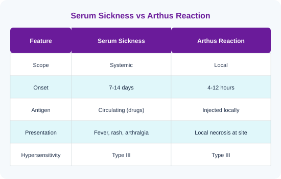
Lupus nephritis biopsy is one of the most tested topics in renal pathology. The WHO/ISN classification assigns six classes. USMLE focuses on Class IV (diffuse proliferative), which is the most common and most severe form.

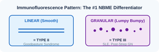
| NBME Trap Alert: Type III • Fever, joint pain, and rash 7 to 14 days after a new drug is SERUM SICKNESS (Type III), not drug allergy (Type I). Timing is the giveaway. • Granular IF = Type III. Linear IF = Type II. This is the single most tested IF distinction. • Low complement (C3, C4) + proteinuria + anti-dsDNA = SLE immune complex nephritis (Type III). • Hypersensitivity pneumonitis = Type III + Type IV combined. Farmer or bird exposure + granulomas + restrictive PFTs. |
Type III = Circulating antigen-antibody immune complexes + Complement C3a/C5a + Neutrophils + Tissue deposition = Vasculitis, nephritis, serum sickness.

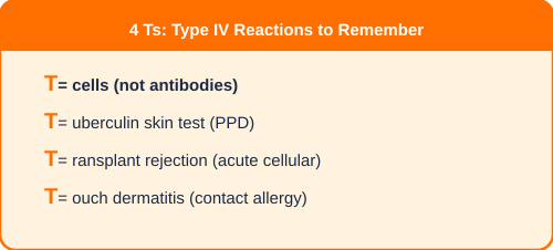
Type IV is the only hypersensitivity reaction that does NOT involve antibodies. It is entirely mediated by T lymphocytes: CD4+ helper T cells (mainly Th1) and CD8+ cytotoxic T cells. The hallmark is delayed onset, requiring 48 to 72 hours after antigen exposure for T cell activation, cytokine production, and macrophage recruitment. If USMLE says “no antibodies involved,” the answer is Type IV.
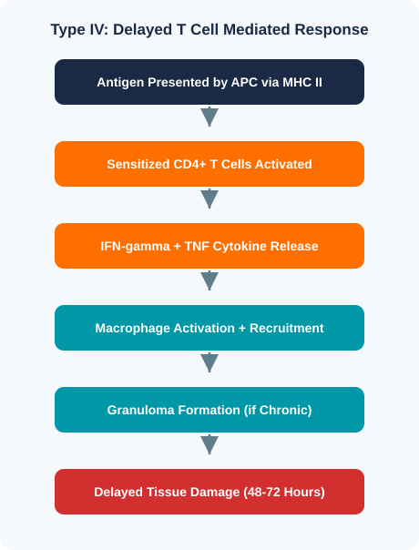
| Subtype | Key Cell | Mechanism | Classic Example |
| IVa (Th1 mediated) | CD4+ Th1 + Macrophage | IFN-gamma activates macrophages | TB granuloma, leprosy, Crohn disease |
| IVb (Th2 mediated) | CD4+ Th2 + Eosinophil | IL-4/IL-5 drive eosinophilia | Chronic allergic asthma, parasitic response |
| IVc (CTL mediated) | CD8+ cytotoxic T cell | Direct killing via perforin/granzyme | Contact dermatitis, graft rejection, SJS/TEN |
| IVd (T cell + Neutrophil) | T cell + Neutrophil | CXCL8 recruits neutrophils | Acute generalized exanthematous pustulosis (AGEP) |
| Disease | Mechanism | Key Clinical Feature |
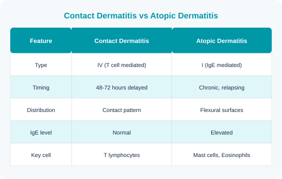
| Contact dermatitis | Sensitized T cells react to hapten on Langerhans cells | Poison ivy, nickel, latex: erythematous vesicular rash 48-72 hrs |
| Tuberculin skin test (PPD) | Memory Th1 cells release IFN-gamma at injection site | Induration (not erythema) measured at 48-72 hours |
| Granulomatous diseases | Chronic macrophage activation by persistent antigens | TB, sarcoidosis, Crohn: epithelioid cells, giant cells, fibrosis |
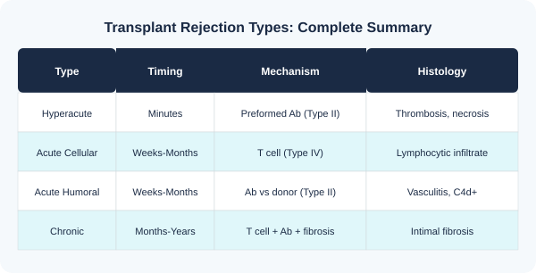
| Acute cellular transplant rejection | Host CD4+ and CD8+ T cells recognize donor MHC | Lymphocytic infiltrate of graft, weeks to months post transplant |
| Chronic transplant rejection | T cell + antibody mediated vascular damage over time | Intimal fibrosis, graft arteriosclerosis, months to years |
| Type 1 diabetes mellitus | CD8+ T cells destroy pancreatic beta cells | Anti-GAD65 and anti-islet cell Ab markers (T cells do damage) |
| Hashimoto thyroiditis | CD8+ T cells + anti-TPO antibodies destroy thyroid | Hypothyroidism, painless goiter, Hurthle cell change |
| Multiple sclerosis | T cells attack CNS myelin (oligodendrocytes) | Relapsing-remitting neurological deficits, periventricular plaques |
| Stevens-Johnson / TEN | CD8+ T cells destroy keratinocytes via Fas/FasL and granulysin | Drug reaction (allopurinol, carbamazepine), epidermal detachment |
| Graft versus host disease | Donor T cells attack host tissues | Post bone marrow transplant: rash, diarrhea, jaundice |
Granulomas are the histologic hallmark of chronic Type IV hypersensitivity. They form when macrophages cannot eliminate the antigen (persistent intracellular pathogens like TB, or non-degradable substances like beryllium). Macrophages transform into epithelioid cells and fuse into multinucleated giant cells. CD4+ T cells surround the granuloma and maintain it via IFN-gamma signaling.
| NBME Trap Alert: Type IV • If the stem says “48 to 72 hours” and describes a rash or induration, the answer is Type IV. No exceptions. • PPD test measures INDURATION (palpable hardening), not erythema. This is a classic wrong answer trap. • Contact dermatitis from poison ivy is Type IV (T cells). Atopic dermatitis is Type I (IgE). Do not be confused. • Type 1 DM is immune mediated (CD8+ T cells destroy beta cells) despite having antibody markers (anti-GAD65). The T cells do the damage. • SJS/TEN is a Type IV drug reaction (CD8+ T cells kill keratinocytes). It is NOT Type I anaphylaxis. |
Type IV = T cells only + No antibodies + Delayed 48-72 hrs + Granulomas + Contact dermatitis + Transplant rejection.
Allergic asthma has two phases. The immediate phase (minutes) is Type I: IgE cross links mast cells, releasing histamine and leukotrienes causing acute bronchoconstriction. The late phase (4 to 8 hours) involves Th2 cells, eosinophils, and chronic inflammation, which is a Type IV component. USMLE tests both phases. Acute wheezing = Type I mediators. Chronic eosinophilic inflammation and airway remodeling = Th2/Type IV.
Before tackling practice questions, study the traps. NBME writers use the same wrong answer designs repeatedly. If you recognize the pattern, you eliminate two to three choices before reading the options.
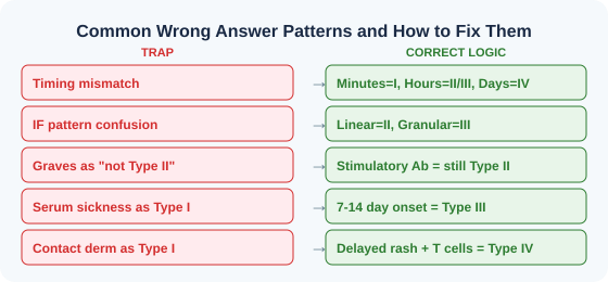
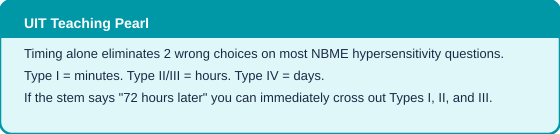
| The 5 Second Rule for Hypersensitivity Questions Step 1: What is the TIMING? Minutes = I. Hours = II/III. Days = IV. Step 2: Are ANTIBODIES involved? Yes = I, II, or III. No = IV. Step 3: Is the target a CELL SURFACE or a SOLUBLE COMPLEX? Cell surface = II. Soluble = III. Step 4: Check the IF pattern if given. Linear = II. Granular = III. This 4 step algorithm solves 90% of NBME hypersensitivity questions in under 30 seconds. |
| HYPERSENSITIVITY REACTIONS: Complete Mindmap Summary | |||
| TYPE IImmediate / Anaphylactic | TYPE IICytotoxic / Antibody Mediated | TYPE IIIImmune Complex | TYPE IVDelayed / T Cell Mediated |
| IgE + Mast Cells | IgG / IgM + Complement | IgG / IgM Complexes | T Cells (CD4+ / CD8+) |
| OnsetMinutes | OnsetHours | OnsetHours to Days | Onset48 to 72 Hours |
| Allergen crosslinks IgE on mast cells causing degranulation and histamine release | Antibodies bind cell surface antigens activating complement or ADCC leading to cell destruction | Antigen antibody complexes deposit in tissues activating complement and recruiting neutrophils | Sensitized T cells release cytokines (CD4+) or directly kill target cells (CD8+) on re exposure |
| AnaphylaxisAllergic asthmaAllergic rhinitisUrticariaFood allergies | Autoimmune hemolytic anemiaGoodpasture syndromeRheumatic feverABO transfusion reactionsHemolytic disease of newborn | SLE (lupus nephritis)Polyarteritis nodosaSerum sicknessArthus reactionPoststreptococcal GN | Contact dermatitisTB skin test (PPD)Transplant rejection (acute)Type 1 diabetes mellitusMultiple sclerosis |
| BOARD MNEMONIC: “ACID” = Anaphylactic / Cytotoxic / Immune Complex / DelayedTiming mnemonic: Type I = minutes | Type II = hours | Type III = hours to days | Type IV = days (48 to 72 hr) |
| Feature | Type I | Type II | Type III | Type IV |
| Antibody | IgE | IgG / IgM | IgG / IgM | None (T cells) |
| Onset | Minutes | Hours | Hours to days | 48 to 72 hours |
| Mediator Cells | Mast cells, basophils | Complement, NK cells, macrophages | Complement, neutrophils | CD4+ T cells, CD8+ T cells, macrophages |
| Effector Mechanism | Mast cell degranulation | Cell lysis, opsonization, ADCC | Complex deposition, complement | Cytokine release, direct cell kill |
| IF Pattern | N/A | Linear | Granular | N/A |
| Complement | Not involved | Activated (classical) | Activated and consumed | Not involved |
| Prototype | Anaphylaxis | Goodpasture | SLE nephritis | Contact dermatitis |
| Key Lab Test | Serum IgE, skin prick | Direct Coombs | Low C3/C4, ANA | PPD / patch test |

A 28 year old woman presents 20 minutes after eating shrimp with throat tightness, diffuse urticaria, and blood pressure 78/50 mmHg. Which mediator is most directly responsible for her hypotension?
Answer: C) Histamine. Anaphylaxis (Type I). IgE mediated mast cell degranulation releases histamine causing vasodilation and increased permeability leading to distributive shock. IFN-gamma is Type IV. IgG/IgM are Types II/III.
A 35 year old man with hemoptysis and hematuria undergoes renal biopsy showing smooth continuous IgG along the glomerular basement membrane on immunofluorescence. Which hypersensitivity type?
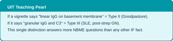
Answer: B) Type II. Linear IF is pathognomonic for Type II, where antibodies target fixed antigens on the basement membrane (Goodpasture). Granular IF would indicate Type III.
A 12 year old boy develops fever, urticaria, arthralgia, and proteinuria 10 days after starting penicillin. What is the mechanism?
Answer: C) Immune complex deposition. Serum sickness (Type III). The 7-14 day onset, systemic symptoms, and drug trigger are classic. Type I would present within minutes.
A nurse develops pruritic erythematous vesicles on both hands 72 hours after wearing new latex gloves. Biopsy shows spongiotic dermatitis with perivascular lymphocytes. This reaction is mediated by:
Answer: D) T lymphocytes. Contact dermatitis is Type IV. The 72 hour timeline, contact pattern, and lymphocytic infiltrate confirm T cell mediation.
A 45 year old woman with Graves disease has elevated free T4, suppressed TSH, and thyroid stimulating immunoglobulins. Which antibody class mediates this?
Answer: C) IgG. Graves is Type II where IgG stimulates the TSH receptor. This is the non-cytotoxic stimulatory subtype of Type II.
A 6 year old presents with periorbital edema, cola colored urine, and hypertension 2 weeks after pharyngitis. Renal biopsy shows subepithelial humps on EM. Expected IF pattern?
Answer: B) Granular IgG and C3. Post-strep GN is Type III. Immune complexes deposit in granular pattern. Subepithelial humps on EM are classic. Linear = Goodpasture (Type II). IgA mesangial = IgA nephropathy (different disease).
A kidney transplant turns blue and must be removed within minutes of anastomosis. Mechanism?
Answer: A) Preformed antibodies. Hyperacute rejection: minutes, preformed antibodies (Type II) attack donor endothelium, activate complement, cause thrombosis. T cell rejection takes weeks.
A 30 year old woman with SLE has anti-dsDNA antibodies, low C3/C4, proteinuria, and wire loop lesions on biopsy. Primary renal pathogenic mechanism?
Answer: C) Immune complex deposition. Lupus nephritis is Type III. Anti-dsDNA immune complexes deposit in glomeruli. Low complement = consumed by activation. Anti-GBM (B) = Goodpasture, not SLE.
A PPD test shows 15 mm induration at 72 hours. Which cytokine is most critical for the observed induration?
Answer: D) IFN-gamma. PPD is classic Type IV. Th1 cells release IFN-gamma activating macrophages causing induration. IL-4/IL-5 are Th2 (Type I). IL-10 is immunosuppressive.
A 22 year old woman has fatigable weakness worsening throughout the day, ptosis, and difficulty chewing. Anti-AChR antibodies detected. Mechanism of weakness?
Answer: C) Antibody mediated receptor blockade. Myasthenia gravis is Type II with a BLOCKING mechanism. IgG blocks AChR and causes receptor internalization. This is the non-cytotoxic functional subtype. Not immune complex (B) or T cell (D).

| Exam Traps: Memorize These Before Walking In • Linear IF = Type II (Goodpasture). Granular IF = Type III (SLE, PSGN). This alone answers many questions. • Serum sickness = 7-14 days after drug. Drug allergy Type I = minutes. Do not be confused. • Graves is Type II but NOT destructive. It is stimulatory. MG is Type II but blocking. • Contact dermatitis = Type IV. Atopic dermatitis = Type I. The word “contact” = delayed = T cells. • Type IV is the ONLY type without antibodies. If “no antibodies” appear in the stem, the answer is Type IV. • Hyperacute rejection = pre formed Ab (minutes). Acute cellular = T cells (weeks). Chronic = both (years). • Low complement in the lab = think Type III (immune complexes consume complement). • Post-strep GN appears 2-3 weeks after pharyngitis. The delay allows immune complex formation. |
Start with timing. Minutes = Type I. Hours = Type II or III. Days = Type IV. Then check antibody involvement. Antibodies against a cell surface = Type II. Immune complex deposition = Type III. No antibodies mentioned and T cells involved = Type IV.
Anaphylaxis is Type I (IgE, mast cells, seconds to minutes). Serum sickness is Type III (immune complexes, 7 to 14 days after exposure). The timing and mechanism are completely different despite both being drug reactions.
Type II classification is based on mechanism (antibody targeting cell surface antigen), not outcome. In Graves, IgG antibodies bind and stimulate the TSH receptor. The target is a cell surface receptor, making it Type II even though the effect is stimulatory rather than destructive.
Yes. Hypersensitivity pneumonitis involves both Type III (immune complex) and Type IV (granulomatous). Chronic asthma starts as Type I (IgE) but develops a Type IV (Th2/eosinophilic) late phase. USMLE may test either phase depending on what the vignette emphasizes.
Linear = Type II. The antibodies target antigens fixed in a continuous line along the basement membrane (like a straight highway). Granular = Type III. Immune complexes deposit randomly in an irregular lumpy bumpy pattern (like scattered gravel). This distinction alone answers the majority of IF questions on NBME.
In Type II, antibodies attack antigens that are fixed to a cell surface or basement membrane, causing tissue specific damage. In Type III, antibodies form soluble complexes with circulating antigens, and these complexes deposit wherever blood flow carries them. Type II damage is predictable (always the same tissue). Type III damage depends on where complexes lodge.
Contact dermatitis involves T cells recognizing haptens presented by Langerhans cells, not IgE mediated mast cell degranulation. The delayed 48-72 hour onset, lymphocytic infiltrate on biopsy, and absence of elevated IgE all confirm T cell mediation. Atopic dermatitis is the IgE mediated (Type I) skin condition.
Transplant rejection involves multiple types. Hyperacute rejection (minutes) is Type II: preformed antibodies attack donor endothelium. Acute cellular rejection (weeks to months) is Type IV: host T cells attack donor MHC. Chronic rejection involves both antibody and T cell mediated mechanisms over months to years.
In Type II, complement activation produces C3b (opsonization) and MAC (cell lysis). In Type III, complement generates C3a and C5a (anaphylatoxins that recruit neutrophils). Low serum C3 and C4 suggest complement consumption by immune complexes, pointing toward Type III diseases like SLE.
Penicillin, cephalosporins, sulfonamides, and anti-thymocyte globulin are the most tested causes. The drug acts as a hapten, forming immune complexes that deposit systemically. Presentation: fever, urticaria, lymphadenopathy, arthralgia, and proteinuria 7 to 14 days after drug exposure.
This guide covers hypersensitivity reactions the way they should be taught: with clinical logic, pattern recognition, and exam awareness. If this approach resonated with you, the UIT Program by IMG Helping Hands goes far deeper across every high yield Step 1 topic.
UIT does not just teach you topics. It teaches you how to think through USMLE vignettes the way high scorers do. Every concept is broken down with clinical integration, NBME pattern analysis, pharmacology links, and rapid review systems designed specifically for IMGs who need efficient, focused preparation.
| What You Get With UIT Mentorship Systems based concept teaching with clinical integration across all organ systems NBME pattern recognition training for every high yield topic Original mnemonics and visual learning frameworks you will not find anywhere else Live mentorship sessions with USMLE experienced faculty who understand IMG challenges Structured study plans tailored for IMG schedules, resources, and learning gaps Pharmacology and pathology integration in every teaching session |
Explore the UIT Program and IMGHH resources at IMG Helping Hands. Join thousands of IMGs who are preparing smarter, not harder.
IMG Helping Hands – UIT Program
Systems-based learning, NBME-focused training, and live mentorship designed specifically for IMGs.
Structured mentorship. Real results.
Strengthen your foundation with these companion resources from IMG Helping Hands:
1. Le T, Bhushan V. First Aid for the USMLE Step 1 2026. McGraw Hill Education.
2. Kumar V, Abbas AK, Aster JC. Robbins and Cotran Pathologic Basis of Disease. 10th Ed. Elsevier.
3. Sattar H. Pathoma: Fundamentals of Pathology.
4. NBME Subject Examination Content Outlines. National Board of Medical Examiners.
5. Abbas AK, Lichtman AH, Pillai S. Cellular and Molecular Immunology. 10th Ed. Elsevier.
6. Janeway CA, Travers P, Walport M. Immunobiology: The Immune System in Health and Disease.
7. UWorld USMLE Step 1 Question Bank Explanations (mechanism-based content alignme
Articles published by IMG Helping Hands are prepared by our team using information from direct experience, publicly available resources, and educational references. AI tools may be used to assist with drafting, proofreading, and formatting; however, all content undergoes review and approval before publication.
The information provided is intended for educational purposes only. Requirements, policies, and processes may change over time. Readers should consult official sources for the most current information.